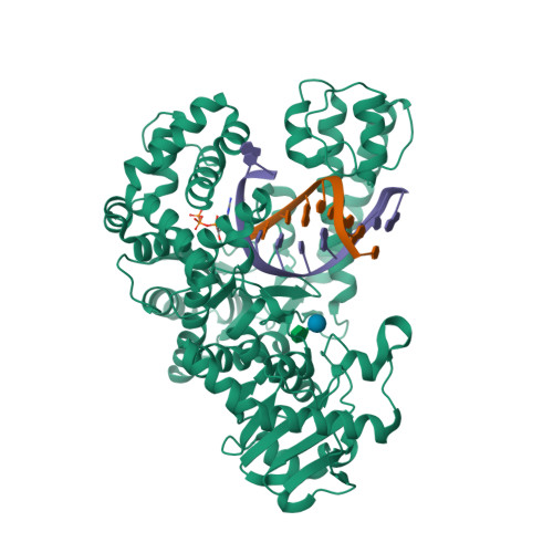
Structural basis for blockage of DNA synthesis by a thymine dimer lesion in a high-fidelity DNA polymerase
Walsh, A.R., Beese, L.S., Wu, E.Y.To be published.
Experimental Data Snapshot
Starting Model: experimental
View more details
Entity ID: 1 | |||||
|---|---|---|---|---|---|
| Molecule | Chains | Sequence Length | Organism | Details | Image |
| DNA polymerase I | A, B [auth D] | 580 | Geobacillus stearothermophilus | Mutation(s): 1 Gene Names: DPO1, polA EC: 2.7.7.7 | 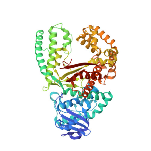 |
UniProt | |||||
Find proteins for D9N168 (Geobacillus stearothermophilus) Explore D9N168 Go to UniProtKB: D9N168 | |||||
Entity Groups | |||||
| Sequence Clusters | 30% Identity50% Identity70% Identity90% Identity95% Identity100% Identity | ||||
| UniProt Group | D9N168 | ||||
Sequence AnnotationsExpand | |||||
| |||||
| Ligands 3 Unique | |||||
|---|---|---|---|---|---|
| ID | Chains | Name / Formula / InChI Key | 2D Diagram | 3D Interactions | |
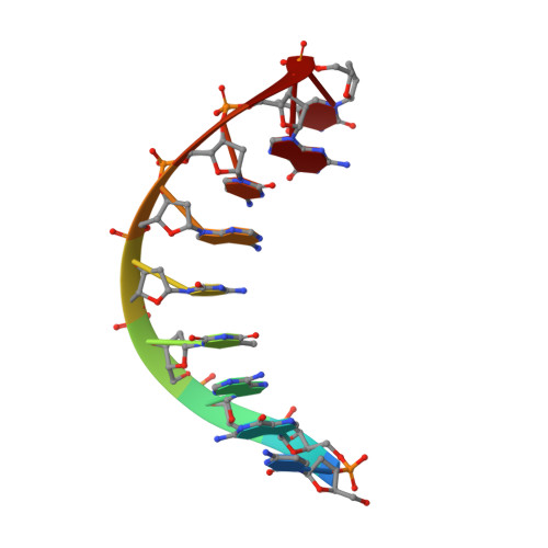
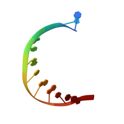
| DTP Query on DTP | I [auth A], J [auth D] | 2'-DEOXYADENOSINE 5'-TRIPHOSPHATE C10 H16 N5 O12 P3 SUYVUBYJARFZHO-RRKCRQDMSA-N |  | ||
| MPD Query on MPD | L [auth D] | (4S)-2-METHYL-2,4-PENTANEDIOL C6 H14 O2 SVTBMSDMJJWYQN-YFKPBYRVSA-N |  | ||
| SO4 Query on SO4 | K [auth D] | SULFATE ION O4 S QAOWNCQODCNURD-UHFFFAOYSA-L |  | ||
Entity ID: 4 | |||||
|---|---|---|---|---|---|
| ID | Chains | Name | Type/Class | 2D Diagram | 3D Interactions |
| PRD_900003 Query on PRD_900003 | G, H | sucrose | Oligosaccharide / Nutrient |  | |
| Length ( Å ) | Angle ( ˚ ) |
|---|---|
| a = 93.875 | α = 90 |
| b = 109.068 | β = 90 |
| c = 150.096 | γ = 90 |
| Software Name | Purpose |
|---|---|
| XDS | data reduction |
| XSCALE | data scaling |
| PHENIX | refinement |
| PDB_EXTRACT | data extraction |
| REFMAC | phasing |
| Funding Organization | Location | Grant Number |
|---|---|---|
| National Institutes of Health/National Institute of General Medical Sciences (NIH/NIGMS) | United States | RO1 GM091487 |
RCSB PDB is hosted by

RCSB PDB is a member of the

All questions and comments will receive a response in a timely manner.