7E44

Crystal structure of NudC complexed with dpCoA


Experimental Data Snapshot

  • Method: X-RAY DIFFRACTION
  • Resolution: 2.00 Å
  • R-Value Free: 0.248 
  • R-Value Work: 0.234 
  • R-Value Observed: 0.235 

wwPDB Validation   3D Report Full Report


Ligand Structure Quality Assessment 


This is version 1.2 of the entry. See complete history


Literature

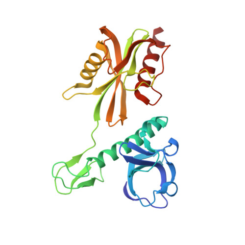
Structural insights into dpCoA-RNA decapping by NudC.

Zhou, W.Guan, Z.Zhao, F.Ye, Y.Yang, F.Yin, P.Zhang, D.

(2021) RNA Biol 18: 244-253

  • DOI: https://doi.org/10.1080/15476286.2021.1936837
  • Primary Citation of Related Structures:  
    7E44

  • PubMed Abstract: 

    Various kinds of cap structures, such as m 7 G, triphosphate groups, NAD and dpCoA, protect the 5' terminus of RNA. The cap structures bond covalently to RNA and affect its stability, translation, and transport. The removal of the caps is mainly executed by Nudix hydrolase family proteins, including Dcp2, RppH and NudC. Numerous efforts have been made to elucidate the mechanism underlying the removal of m 7 G, triphosphate group, and NAD caps. In contrast, few studies related to the cleavage of the RNA dpCoA cap have been conducted. Here, we report the hydrolytic activity of Escherichia coli NudC towards dpCoA and dpCoA-capped RNA in vitro . We also determined the crystal structure of dimeric NudC in complex with dpCoA at 2.0 Å resolution. Structural analysis revealed that dpCoA is recognized and hydrolysed in a manner similar to NAD. In addition, NudC may also remove other dinucleotide derivative caps of RNA, which comprise the AMP moieties. NudC homologs in Saccharomyces cerevisiae and Arabidopsis thaliana exhibited similar dpCoA decapping (deCoAping) activity. These results together indicate a conserved mechanism underpinning the hydrolysis of dpCoA-capped RNA in both prokaryotes and eukaryotes.


  • Organizational Affiliation

    National Key Laboratory of Crop Genetic Improvement, Hubei Hongshan Laboratory, Huazhong Agricultural University, Wuhan, China.


Macromolecules
Find similar proteins by:  (by identity cutoff)  |  3D Structure
Entity ID: 1
MoleculeChains Sequence LengthOrganismDetailsImage
NADH pyrophosphataseA,
B,
C [auth E],
D [auth F]
259Escherichia coli BL21(DE3)Mutation(s): 0 
Gene Names: nudCECBD_4036HO396_19860
EC: 3.6.1.22
UniProt
Find proteins for P32664 (Escherichia coli (strain K12))
Explore P32664 
Go to UniProtKB:  P32664
Entity Groups  
Sequence Clusters30% Identity50% Identity70% Identity90% Identity95% Identity100% Identity
UniProt GroupP32664
Sequence Annotations
Expand
  • Reference Sequence
Experimental Data & Validation

Experimental Data

  • Method: X-RAY DIFFRACTION
  • Resolution: 2.00 Å
  • R-Value Free: 0.248 
  • R-Value Work: 0.234 
  • R-Value Observed: 0.235 
  • Space Group: P 1
Unit Cell:
Length ( Å )Angle ( ˚ )
a = 60.837α = 97.243
b = 61.651β = 100.386
c = 93.893γ = 111.139
Software Package:
Software NamePurpose
PHENIXrefinement
DIALSdata reduction
Aimlessdata scaling
PHASERphasing

Structure Validation

View Full Validation Report


Ligand Structure Quality Assessment 


Entry History 

Deposition Data

Revision History  (Full details and data files)

  • Version 1.0: 2021-07-28
    Type: Initial release
  • Version 1.1: 2022-01-26
    Changes: Database references
  • Version 1.2: 2023-11-29
    Changes: Data collection, Refinement description