7T6I

Crystal structure of HLA-DP1 in complex with pp65 peptide in reverse orientation


Experimental Data Snapshot

  • Method: X-RAY DIFFRACTION
  • Resolution: 2.30 Å
  • R-Value Free: 
    0.224 (Depositor), 0.230 (DCC) 
  • R-Value Work: 
    0.181 (Depositor), 0.180 (DCC) 
  • R-Value Observed: 
    0.183 (Depositor) 

Starting Model: experimental
View more details

wwPDB Validation   3D Report Full Report


Ligand Structure Quality Assessment 

Created with Raphaël 2.3.0Worse 01 BetterLigand structure goodness of fit to experimental dataBest fitted NAGClick on this verticalbar to view details

This is version 1.2 of the entry. See complete history


Literature

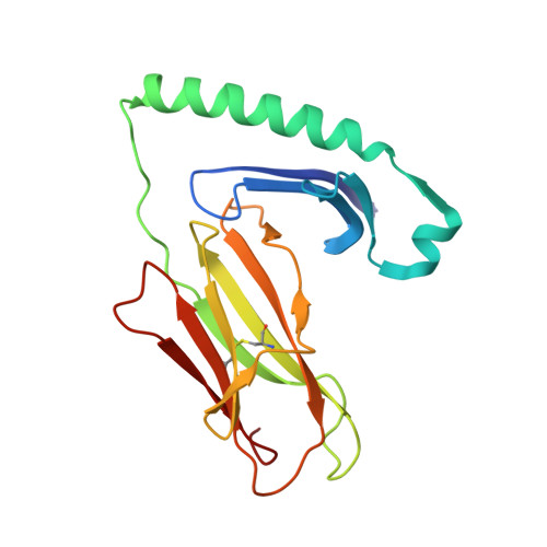
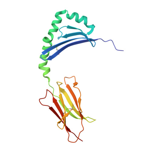
Human T cells recognize HLA-DP-bound peptides in two orientations.

Klobuch, S.Lim, J.J.van Balen, P.Kester, M.G.D.de Klerk, W.de Ru, A.H.Pothast, C.R.Jedema, I.Drijfhout, J.W.Rossjohn, J.Reid, H.H.van Veelen, P.A.Falkenburg, J.H.F.Heemskerk, M.H.M.

(2022) Proc Natl Acad Sci U S A 119: e2214331119-e2214331119

  • DOI: https://doi.org/10.1073/pnas.2214331119
  • Primary Citation of Related Structures:  
    7T6I

  • PubMed Abstract: 

    Human leukocyte antigen (HLA) molecules present small peptide antigens to T cells, thereby allowing them to recognize pathogen-infected and cancer cells. A central dogma over the last 50+ y is that peptide binding to HLA molecules is mediated by the docking of side chains of particular amino acids in the peptide into pockets in the HLA molecules in a conserved N- to C-terminal orientation. Whether peptides can be presented in a reversed C- to N-terminal orientation remains unclear. Here, we performed large-scale identification of peptides bound to HLA-DP molecules and observed that in addition to peptide binding in an N- to C-terminal orientation, in 9 out of 14 HLA-DP allotypes, reverse motifs are found, compatible with C- to N-terminal peptide binding. Moreover, we isolated high-avidity human cytomegalovirus (CMV)-specific HLA-DP-restricted CD4 + T cells from the memory repertoire of healthy donors and demonstrate that such T cells recognized CMV-derived peptides bound to HLA-DPB1*01:01 or *05:01 in a reverse C- to N-terminal manner. Finally, we obtained a high-resolution HLA-DPB1*01:01-CMVpp65 (142-158) peptide crystal structure, which is the molecular basis for C- to N-terminal peptide binding to HLA-DP. Our results point to unique features of HLA-DP molecules that substantially broaden the HLA class II bound peptide repertoire to combat pathogens and eliminate cancer cells.


  • Organizational Affiliation

    Department of Hematology, Leiden University Medical Center, 2333 ZA Leiden, The Netherlands.


Macromolecules
Find similar proteins by:  (by identity cutoff)  |  3D Structure
Entity ID: 1
MoleculeChains Sequence LengthOrganismDetailsImage
HLA class II histocompatibility antigen DP alpha chain183Homo sapiensMutation(s): 0 
Gene Names: HLA-DPA1
UniProt
Find proteins for Q95HB9 (Homo sapiens)
Explore Q95HB9 
Go to UniProtKB:  Q95HB9
Entity Groups  
Sequence Clusters30% Identity50% Identity70% Identity90% Identity95% Identity100% Identity
UniProt GroupQ95HB9
Glycosylation
Glycosylation Sites: 2
Sequence Annotations
Expand
  • Reference Sequence
Find similar proteins by:  (by identity cutoff)  |  3D Structure
Entity ID: 2
MoleculeChains Sequence LengthOrganismDetailsImage
MHC class II antigen191Homo sapiensMutation(s): 0 
Gene Names: HLA-DPB1DPB1
UniProt
Find proteins for S6B6U4 (Homo sapiens)
Explore S6B6U4 
Go to UniProtKB:  S6B6U4
Entity Groups  
Sequence Clusters30% Identity50% Identity70% Identity90% Identity95% Identity100% Identity
UniProt GroupS6B6U4
Glycosylation
Glycosylation Sites: 1
Sequence Annotations
Expand
  • Reference Sequence

Find similar proteins by:  Sequence   |   3D Structure  

Entity ID: 3
MoleculeChains Sequence LengthOrganismDetailsImage
pp65 peptide17Human herpesvirus 5 strain AD169Mutation(s): 0 
UniProt
Find proteins for P06725 (Human cytomegalovirus (strain AD169))
Explore P06725 
Go to UniProtKB:  P06725
Entity Groups  
Sequence Clusters30% Identity50% Identity70% Identity90% Identity95% Identity100% Identity
UniProt GroupP06725
Sequence Annotations
Expand
  • Reference Sequence
Experimental Data & Validation

Experimental Data

  • Method: X-RAY DIFFRACTION
  • Resolution: 2.30 Å
  • R-Value Free:  0.224 (Depositor), 0.230 (DCC) 
  • R-Value Work:  0.181 (Depositor), 0.180 (DCC) 
  • R-Value Observed: 0.183 (Depositor) 
Space Group: C 1 2 1
Unit Cell:
Length ( Å )Angle ( ˚ )
a = 112.36α = 90
b = 62.364β = 91.523
c = 72.796γ = 90
Software Package:
Software NamePurpose
PHENIXrefinement
XDSdata reduction
Aimlessdata scaling
PHASERphasing

Structure Validation

View Full Validation Report


Ligand Structure Quality Assessment 

Created with Raphaël 2.3.0Worse 01 BetterLigand structure goodness of fit to experimental dataBest fitted NAGClick on this verticalbar to view details

Entry History & Funding Information

Deposition Data


Funding OrganizationLocationGrant Number
Australian Research Council (ARC)Australia--

Revision History  (Full details and data files)

  • Version 1.0: 2022-12-07
    Type: Initial release
  • Version 1.1: 2023-10-25
    Changes: Data collection, Refinement description
  • Version 1.2: 2024-11-13
    Changes: Structure summary