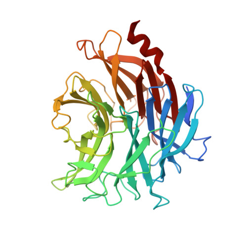
8UB5

The Apo NanH structure from Clostridium perfringens


Experimental Data Snapshot

  • Method: X-RAY DIFFRACTION
  • Resolution: 1.80 Å
  • R-Value Free: 0.197 
  • R-Value Work: 0.164 
  • R-Value Observed: 0.165 

Starting Model: experimental
View more details

wwPDB Validation   3D Report Full Report


This is version 1.2 of the entry. See complete history


Literature

A "terminal" case of glycan catabolism: Structural and enzymatic characterization of the sialidases of Clostridium perfringens.

Medley, B.J.Low, K.E.Irungu, J.D.W.Kipchumba, L.Daneshgar, P.Liu, L.Garber, J.M.Klassen, L.Inglis, G.D.Boons, G.J.Zandberg, W.F.Abbott, D.W.Boraston, A.B.

(2024) J Biol Chem 300: 107750-107750

  • DOI: https://doi.org/10.1016/j.jbc.2024.107750
  • Primary Citation of Related Structures:  
    8U2A, 8U5O, 8UB5, 8UL7, 8ULE, 8UM0, 8UVV

  • PubMed Abstract: 

    Sialic acids are commonly found on the terminal ends of biologically important carbohydrates, including intestinal mucin O-linked glycans. Pathogens such as Clostridium perfringens, the causative agent of necrotic enteritis (NE) in poultry and humans, have the ability to degrade host mucins and colonize the mucus layer, which involves removal of the terminal sialic acid by carbohydrate-active enzymes (CAZymes). Here we present the structural and biochemical characterization of the GH33 catalytic domains of the three sialidases of C. perfringens and probe their substrate specificity. The catalytically active domains, which we refer to as NanH GH33 , NanJ GH33 , and NanI GH33 , displayed differential activity on various naturally occurring forms of sialic acid. We report the X-ray crystal structures of these domains in complex with relevant sialic acid variants revealing the molecular basis of how each catalytic domain accommodates different sialic acids. NanH GH33 displays a distinct preference for α-2,3-linked sialic acid, but can process α-2,6-linked sialic acid. NanJ GH33 and NanI GH33 both exhibit the ability to process α-2,3- and α-2,6-linked sialic acid without any significant apparent preference. All three enzymes were sensitive to generic and commercially available sialidase inhibitors, which impeded sialidase activity in cultures as well as the growth of C. perfringens on sialylated glycans. The knowledge gained in these studies can be applied to in vivo models for C. perfringens growth and metabolism of mucin O-glycans, with a view towards future mitigation of bacterial colonization and infection of intestinal tissues.


  • Organizational Affiliation

    Department of Biochemistry & Microbiology, University of Victoria, Victoria, BC, V8P 5C2 Canada.


Macromolecules
Find similar proteins by:  (by identity cutoff)  |  3D Structure
Entity ID: 1
MoleculeChains Sequence LengthOrganismDetailsImage
Sialidase383Clostridium perfringens ATCC 13124Mutation(s): 0 
Gene Names: nanH
EC: 3.2.1.18
UniProt
Find proteins for B1V1R3 (Clostridium perfringens D str. JGS1721)
Explore B1V1R3 
Go to UniProtKB:  B1V1R3
Entity Groups  
Sequence Clusters30% Identity50% Identity70% Identity90% Identity95% Identity100% Identity
UniProt GroupB1V1R3
Sequence Annotations
Expand
  • Reference Sequence
Small Molecules
Ligands 2 Unique
IDChains Name / Formula / InChI Key2D Diagram3D Interactions
EDO
Query on EDO

Download Ideal Coordinates CCD File 
B [auth A]
C [auth A]
D [auth A]
E [auth A]
F [auth A]
B [auth A],
C [auth A],
D [auth A],
E [auth A],
F [auth A],
G [auth A],
H [auth A],
I [auth A],
J [auth A],
K [auth A],
L [auth A],
M [auth A],
N [auth A],
O [auth A]
1,2-ETHANEDIOL
C2 H6 O2
LYCAIKOWRPUZTN-UHFFFAOYSA-N
ACE
Query on ACE

Download Ideal Coordinates CCD File 
AA [auth A]
BA [auth A]
CA [auth A]
P [auth A]
Q [auth A]
AA [auth A],
BA [auth A],
CA [auth A],
P [auth A],
Q [auth A],
R [auth A],
S [auth A],
T [auth A],
U [auth A],
V [auth A],
W [auth A],
X [auth A],
Y [auth A],
Z [auth A]
ACETYL GROUP
C2 H4 O
IKHGUXGNUITLKF-UHFFFAOYSA-N
Experimental Data & Validation

Experimental Data

  • Method: X-RAY DIFFRACTION
  • Resolution: 1.80 Å
  • R-Value Free: 0.197 
  • R-Value Work: 0.164 
  • R-Value Observed: 0.165 
  • Space Group: C 1 2 1
Unit Cell:
Length ( Å )Angle ( ˚ )
a = 93.436α = 90
b = 64.492β = 102.71
c = 65.747γ = 90
Software Package:
Software NamePurpose
PHENIXrefinement
PHASERphasing
HKL-2000data collection
HKL-2000data reduction

Structure Validation

View Full Validation Report


Entry History & Funding Information

Deposition Data


Funding OrganizationLocationGrant Number
Canadian Glycomics Network (GLYCONET)Canada--

Revision History  (Full details and data files)

  • Version 1.0: 2024-09-04
    Type: Initial release
  • Version 1.1: 2024-09-25
    Changes: Database references, Refinement description
  • Version 1.2: 2024-10-30
    Changes: Database references, Refinement description, Structure summary