RCSB PDB - 9F39: Crystal structure of SARS-CoV-2 Mpro in complex with RK-54

 9F39

Crystal structure of SARS-CoV-2 Mpro in complex with RK-54


Experimental Data Snapshot

  • Method: X-RAY DIFFRACTION
  • Resolution: 2.45 Å
  • R-Value Free: 0.305 
  • R-Value Work: 0.216 

Starting Model: experimental
View more details

wwPDB Validation   3D Report Full Report


Ligand Structure Quality Assessment 

Created with Raphaël 2.3.0Worse 01 BetterLigand structure goodness of fit to experimental dataBest fitted A1H9YClick on this verticalbar to view details

This is version 1.0 of the entry. See complete history


Literature

Structure-Based Optimization of Pyridone alpha-Ketoamides as Inhibitors of the SARS-CoV-2 Main Protease.

Akula, R.K.El Kilani, H.Metzen, A.Roske, J.Zhang, K.Gohl, M.Arisetti, N.Marsh, G.P.Maple, H.J.Cooper, M.S.Karadogan, B.Jochmans, D.Neyts, J.Rox, K.Hilgenfeld, R.Bronstrup, M.

(2025) J Med Chem 

  • DOI: https://doi.org/10.1021/acs.jmedchem.4c02172
  • Primary Citation of Related Structures:  
    8AIU, 8AIV, 8AIZ, 8AJ0, 8AJ1, 9F2V, 9F2X, 9F39, 9F3A, 9FHQ, 9GMQ

  • PubMed Abstract: 

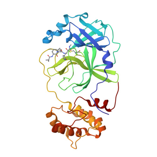
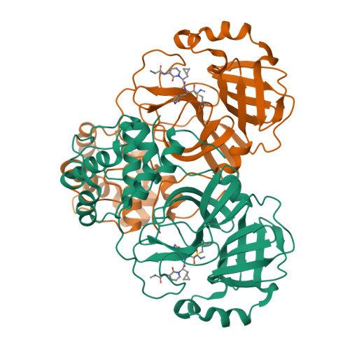
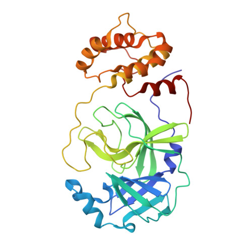
    The main protease M pro is a clinically validated target to treat infections by the coronavirus SARS-CoV-2. Among the first reported M pro inhibitors was the peptidomimetic α-ketoamide 13b , whose cocrystal structure with M pro paved the way for multiple lead-finding studies. We established structure-activity relationships for the 13b series by modifying residues at the P1', P3, and P4 sites. Guided by cocrystal structures, we reduced the P1' substituent size to better fill the pocket and added a fluorine substituent to the pyridone ring, enabling a new hydrogen bond with Gln189 in P3. Among 22 novel analogues, 6d and 12d inhibited M pro with IC 50 s of 110 nM and 40 nM, improving the potency of 13b by up to 9.5-fold. Compound 6d had pronounced antiviral activity with an EC 50 of 1.6 μM and was stable in plasma and microsomes. The study illustrates the potential of structure-based design to systematically improve peptidomimetic α-ketoamides.


  • Organizational Affiliation

    Department of Chemical Biology, Helmholtz Centre for Infection Research, Inhoffenstr. 7, Braunschweig 38124, Germany.


Macromolecules
Find similar proteins by:  (by identity cutoff)  |  3D Structure
Entity ID: 1
MoleculeChains Sequence LengthOrganismDetailsImage
3C-like proteinase nsp5305Severe acute respiratory syndrome coronavirus 2Mutation(s): 0 
Gene Names: rep1a-1b
EC: 3.4.22.69
UniProt
Find proteins for P0DTD1 (Severe acute respiratory syndrome coronavirus 2)
Explore P0DTD1 
Go to UniProtKB:  P0DTD1
Entity Groups  
Sequence Clusters30% Identity50% Identity70% Identity90% Identity95% Identity100% Identity
UniProt GroupP0DTD1
Sequence Annotations
Expand
  • Reference Sequence
Small Molecules
Ligands 1 Unique
IDChains Name / Formula / InChI Key2D Diagram3D Interactions
A1H9Y (Subject of Investigation/LOI)
Query on A1H9Y

Download Ideal Coordinates CCD File 
B [auth A](2R,3R)-3-[[(2S)-3-cyclopropyl-2-[3-(2-methylpropanoylamino)-2-oxidanylidene-pyridin-1-yl]propanoyl]amino]-N-methyl-2-oxidanyl-4-[(3S)-2-oxidanylidenepyrrolidin-3-yl]butanamide
C24 H35 N5 O6
XBTRGWYUGIHXFR-WZGPROSRSA-N
Experimental Data & Validation

Experimental Data

  • Method: X-RAY DIFFRACTION
  • Resolution: 2.45 Å
  • R-Value Free: 0.305 
  • R-Value Work: 0.216 
  • Space Group: C 1 2 1
Unit Cell:
Length ( Å )Angle ( ˚ )
a = 113.702α = 90
b = 53.197β = 102.445
c = 44.856γ = 90
Software Package:
Software NamePurpose
REFMACrefinement
XDSdata reduction
Aimlessdata scaling
MOLREPphasing

Structure Validation

View Full Validation Report


Ligand Structure Quality Assessment 

Created with Raphaël 2.3.0Worse 01 BetterLigand structure goodness of fit to experimental dataBest fitted A1H9YClick on this verticalbar to view details

Entry History & Funding Information

Deposition Data


Funding OrganizationLocationGrant Number
European CommissionEuropean Union--

Revision History  (Full details and data files)

  • Version 1.0: 2025-01-29
    Type: Initial release