3WUR

Structure of DMP19 Complex with 18-crown-6


Experimental Data Snapshot

  • Method: X-RAY DIFFRACTION
  • Resolution: 1.45 Å
  • R-Value Free: 0.204 
  • R-Value Work: 0.169 
  • R-Value Observed: 0.170 

wwPDB Validation   3D Report Full Report


Ligand Structure Quality Assessment 


This is version 1.1 of the entry. See complete history


Literature

Crowning proteins: modulating the protein surface properties using crown ethers.

Lee, C.C.Maestre-Reyna, M.Hsu, K.C.Wang, H.C.Liu, C.I.Jeng, W.Y.Lin, L.L.Wood, R.Chou, C.C.Yang, J.M.Wang, A.H.

(2014) Angew Chem Int Ed Engl 53: 13054-13058

  • DOI: https://doi.org/10.1002/anie.201405664
  • Primary Citation of Related Structures:  
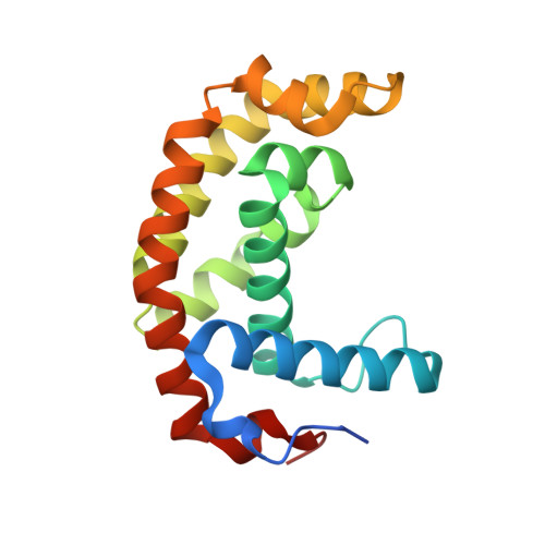
    3WH0, 3WHM, 3WUR

  • PubMed Abstract: 

    Crown ethers are small, cyclic polyethers that have found wide-spread use in phase-transfer catalysis and, to a certain degree, in protein chemistry. Crown ethers readily bind metallic and organic cations, including positively charged amino acid side chains. We elucidated the crystal structures of several protein-crown ether co-crystals grown in the presence of 18-crown-6. We then employed biophysical methods and molecular dynamics simulations to compare these complexes with the corresponding apoproteins and with similar complexes with ring-shaped low-molecular-weight polyethylene glycols. Our studies show that crown ethers can modify protein surface behavior dramatically by stabilizing either intra- or intermolecular interactions. Consequently, we propose that crown ethers can be used to modulate a wide variety of protein surface behaviors, such as oligomerization, domain-domain interactions, stabilization in organic solvents, and crystallization.


  • Organizational Affiliation

    Institute of Biological Chemistry, Academia Sinica, 128 Academia Road, Section 2, Nankang, Taipei 11529 (Taiwan); Core Facilities for Protein Structural Analysis, Academia Sinica, 128 Academia Road, Section 2, Nankang, Taipei 11529 (Taiwan).


Macromolecules
Find similar proteins by:  (by identity cutoff)  |  3D Structure
Entity ID: 1
MoleculeChains Sequence LengthOrganismDetailsImage
Uncharacterized protein
A, B
171Neisseria meningitidis MC58Mutation(s): 0 
Gene Names: NMB0541
UniProt
Find proteins for Q9K0P4 (Neisseria meningitidis serogroup B (strain MC58))
Explore Q9K0P4 
Go to UniProtKB:  Q9K0P4
Entity Groups  
Sequence Clusters30% Identity50% Identity70% Identity90% Identity95% Identity100% Identity
UniProt GroupQ9K0P4
Sequence Annotations
Expand
  • Reference Sequence
Small Molecules
Ligands 4 Unique
IDChains Name / Formula / InChI Key2D Diagram3D Interactions
O4B
Query on O4B

Download Ideal Coordinates CCD File 
C [auth A]
D [auth A]
G [auth B]
H [auth B]
I [auth B]
C [auth A],
D [auth A],
G [auth B],
H [auth B],
I [auth B],
J [auth B]
1,4,7,10,13,16-HEXAOXACYCLOOCTADECANE
C12 H24 O6
XEZNGIUYQVAUSS-UHFFFAOYSA-N
TLA
Query on TLA

Download Ideal Coordinates CCD File 
M [auth B]L(+)-TARTARIC ACID
C4 H6 O6
FEWJPZIEWOKRBE-JCYAYHJZSA-N
SO4
Query on SO4

Download Ideal Coordinates CCD File 
F [auth A],
L [auth B]
SULFATE ION
O4 S
QAOWNCQODCNURD-UHFFFAOYSA-L
EDO
Query on EDO

Download Ideal Coordinates CCD File 
E [auth A],
K [auth B]
1,2-ETHANEDIOL
C2 H6 O2
LYCAIKOWRPUZTN-UHFFFAOYSA-N
Experimental Data & Validation

Experimental Data

  • Method: X-RAY DIFFRACTION
  • Resolution: 1.45 Å
  • R-Value Free: 0.204 
  • R-Value Work: 0.169 
  • R-Value Observed: 0.170 
  • Space Group: P 41 21 2
Unit Cell:
Length ( Å )Angle ( ˚ )
a = 67.1α = 90
b = 67.1β = 90
c = 176.91γ = 90
Software Package:
Software NamePurpose
HKL-2000data collection
PHENIXmodel building
REFMACrefinement
HKL-2000data reduction
HKL-2000data scaling
PHENIXphasing

Structure Validation

View Full Validation Report


Ligand Structure Quality Assessment 


Entry History 

Deposition Data

Revision History  (Full details and data files)

  • Version 1.0: 2014-10-15
    Type: Initial release
  • Version 1.1: 2022-08-24
    Changes: Database references, Derived calculations