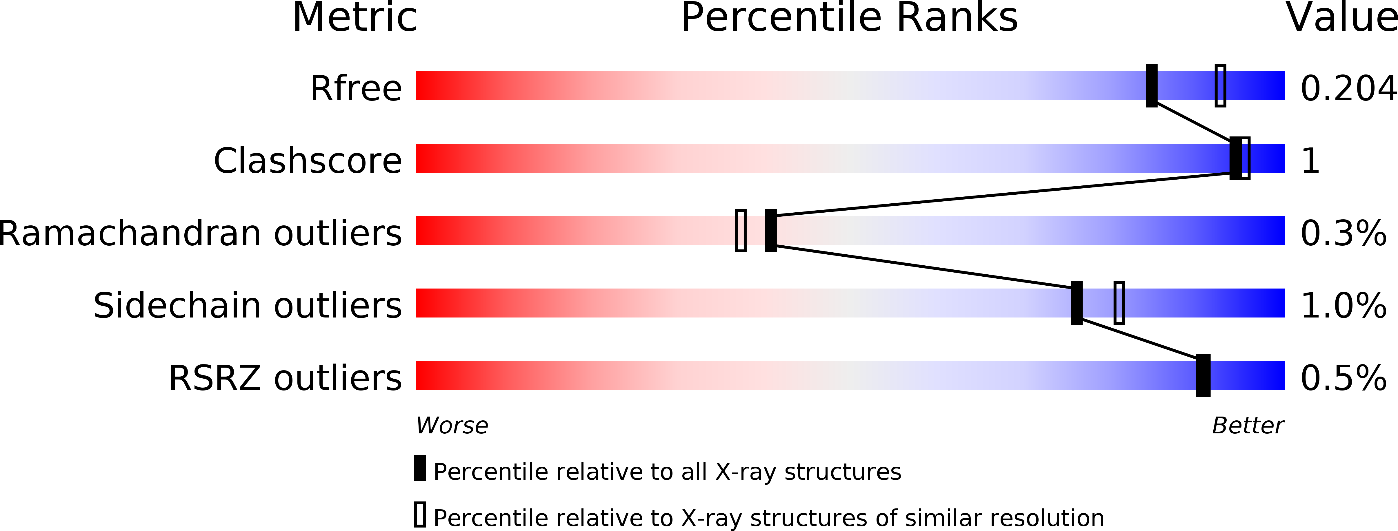
4W4Q

Glucose isomerase structure determined by serial femtosecond crystallography at SACLA


Experimental Data Snapshot

  • Method: X-RAY DIFFRACTION
  • Resolution: 2.00 Å
  • R-Value Free: 0.196 
  • R-Value Work: 0.159 
  • R-Value Observed: 0.161 

wwPDB Validation   3D Report Full Report


This is version 1.4 of the entry. See complete history


Literature

Grease matrix as a versatile carrier of proteins for serial crystallography

Sugahara, M.Mizohata, E.Nango, E.Suzuki, M.Tanaka, T.Masuda, T.Tanaka, R.Shimamura, T.Tanaka, Y.Suno, C.Ihara, K.Pan, D.Kakinouchi, K.Sugiyama, S.Murata, M.Inoue, T.Tono, K.Song, C.Park, J.Kameshima, T.Hatsui, T.Joti, Y.Yabashi, M.Iwata, S.

(2015) Nat Methods 12: 61-63

  • DOI: https://doi.org/10.1038/nmeth.3172
  • Primary Citation of Related Structures:  
    3WUL, 3WUM, 3WXQ, 3WXS, 3WXT, 3WXU, 4W4Q

  • PubMed Abstract: 

    Serial femtosecond X-ray crystallography (SFX) has revolutionized atomic-resolution structural investigation by expanding applicability to micrometer-sized protein crystals, even at room temperature, and by enabling dynamics studies. However, reliable crystal-carrying media for SFX are lacking. Here we introduce a grease-matrix carrier for protein microcrystals and obtain the structures of lysozyme, glucose isomerase, thaumatin and fatty acid-binding protein type 3 under ambient conditions at a resolution of or finer than 2 Å.


  • Organizational Affiliation

    RIKEN SPring-8 Center, Sayo, Japan.


Macromolecules
Find similar proteins by:  (by identity cutoff)  |  3D Structure
Entity ID: 1
MoleculeChains Sequence LengthOrganismDetailsImage
Xylose isomerase388Streptomyces rubiginosusMutation(s): 0 
EC: 5.3.1.5
UniProt
Find proteins for P24300 (Streptomyces rubiginosus)
Explore P24300 
Go to UniProtKB:  P24300
Entity Groups  
Sequence Clusters30% Identity50% Identity70% Identity90% Identity95% Identity100% Identity
UniProt GroupP24300
Sequence Annotations
Expand
  • Reference Sequence
Small Molecules
Ligands 1 Unique
IDChains Name / Formula / InChI Key2D Diagram3D Interactions
CA
Query on CA

Download Ideal Coordinates CCD File 
B [auth A]CALCIUM ION
Ca
BHPQYMZQTOCNFJ-UHFFFAOYSA-N
Experimental Data & Validation

Experimental Data

  • Method: X-RAY DIFFRACTION
  • Resolution: 2.00 Å
  • R-Value Free: 0.196 
  • R-Value Work: 0.159 
  • R-Value Observed: 0.161 
  • Space Group: I 2 2 2
Unit Cell:
Length ( Å )Angle ( ˚ )
a = 94α = 90
b = 100β = 90
c = 103γ = 90
Software Package:
Software NamePurpose
REFMACrefinement

Structure Validation

View Full Validation Report


Entry History & Funding Information

Deposition Data


Funding OrganizationLocationGrant Number
MEXTJapan--

Revision History  (Full details and data files)

  • Version 1.0: 2014-11-05
    Type: Initial release
  • Version 1.1: 2014-12-17
    Changes: Database references
  • Version 1.2: 2015-01-14
    Changes: Database references
  • Version 1.3: 2018-01-24
    Changes: Data collection, Derived calculations, Other, Source and taxonomy
  • Version 1.4: 2024-03-20
    Changes: Data collection, Database references