5U7M

Crystal Structure of HIV-1 BG505 SOSIP.664 Prefusion Env Trimer Bound to Small Molecule HIV-1 Entry Inhibitor BMS-378806 in Complex with Human Antibodies PGT122 and 35O22 at 3.8 Angstrom


Experimental Data Snapshot

  • Method: X-RAY DIFFRACTION
  • Resolution: 3.02 Å
  • R-Value Free: 0.289 
  • R-Value Work: 0.245 
  • R-Value Observed: 0.248 

wwPDB Validation   3D Report Full Report


Ligand Structure Quality Assessment 


This is version 2.0 of the entry. See complete history


Literature

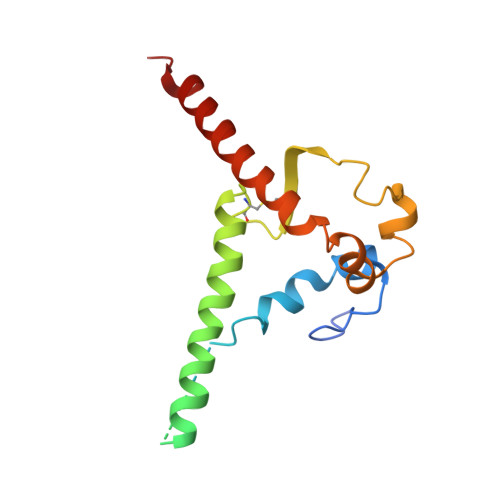
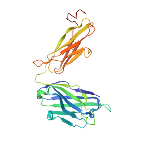
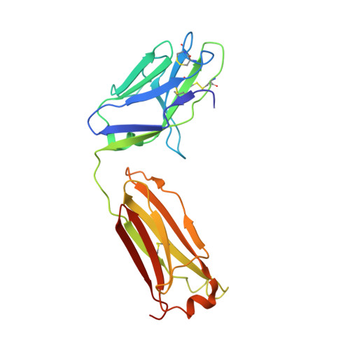
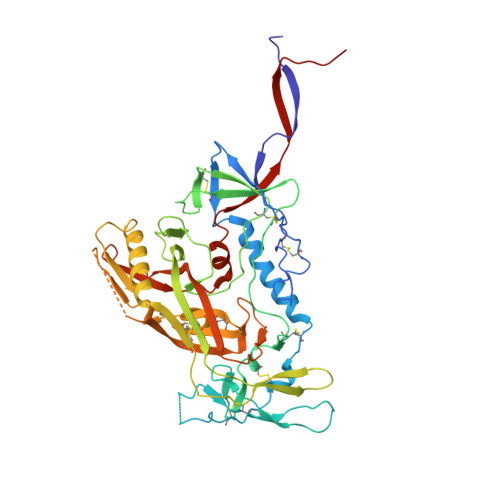
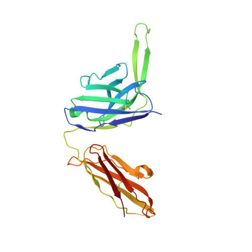
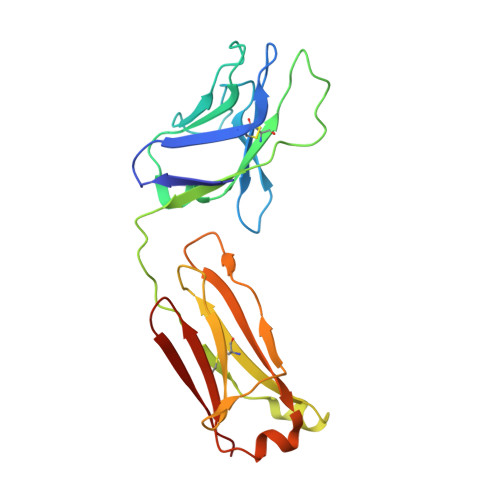
Crystal structures of trimeric HIV envelope with entry inhibitors BMS-378806 and BMS-626529.

Pancera, M.Lai, Y.T.Bylund, T.Druz, A.Narpala, S.O'Dell, S.Schon, A.Bailer, R.T.Chuang, G.Y.Geng, H.Louder, M.K.Rawi, R.Soumana, D.I.Finzi, A.Herschhorn, A.Madani, N.Sodroski, J.Freire, E.Langley, D.R.Mascola, J.R.McDermott, A.B.Kwong, P.D.

(2017) Nat Chem Biol 13: 1115-1122

  • DOI: https://doi.org/10.1038/nchembio.2460
  • Primary Citation of Related Structures:  
    5U7M, 5U7O

  • PubMed Abstract: 

    The HIV-1 envelope (Env) spike is a conformational machine that transitions between prefusion (closed, CD4- and CCR5-bound) and postfusion states to facilitate HIV-1 entry into cells. Although the prefusion closed conformation is a potential target for inhibition, development of small-molecule leads has been stymied by difficulties in obtaining structural information. Here, we report crystal structures at 3.8-Å resolution of an HIV-1-Env trimer with BMS-378806 and a derivative BMS-626529 for which a prodrug version is currently in Phase III clinical trials. Both lead candidates recognized an induced binding pocket that was mostly excluded from solvent and comprised of Env elements from a conserved helix and the β20-21 hairpin. In both structures, the β20-21 region assumed a conformation distinct from prefusion-closed and CD4-bound states. Together with biophysical and antigenicity characterizations, the structures illuminate the allosteric and competitive mechanisms by which these small-molecule leads inhibit CD4-induced structural changes in Env.


  • Organizational Affiliation

    Vaccine Research Center, National Institute of Allergy and Infectious Diseases, National Institutes of Health, Bethesda, Maryland, USA.


Macromolecules
Find similar proteins by:  (by identity cutoff)  |  3D Structure
Entity ID: 1
MoleculeChains Sequence LengthOrganismDetailsImage
Envelope glycoprotein gp160A [auth B]153Human immunodeficiency virus 1Mutation(s): 2 
Gene Names: env
UniProt
Find proteins for Q2N0S5 (Human immunodeficiency virus 1)
Explore Q2N0S5 
Go to UniProtKB:  Q2N0S5
Entity Groups  
Sequence Clusters30% Identity50% Identity70% Identity90% Identity95% Identity100% Identity
UniProt GroupQ2N0S5
Sequence Annotations
Expand
  • Reference Sequence
Find similar proteins by:  (by identity cutoff)  |  3D Structure
Entity ID: 2
MoleculeChains Sequence LengthOrganismDetailsImage
35O22 FAB HEAVY CHAINB [auth D]243Homo sapiensMutation(s): 0 
Entity Groups  
Sequence Clusters30% Identity50% Identity70% Identity90% Identity95% Identity100% Identity
Sequence Annotations
Expand
  • Reference Sequence
Find similar proteins by:  (by identity cutoff)  |  3D Structure
Entity ID: 3
MoleculeChains Sequence LengthOrganismDetailsImage
35O22 FAB LIGHT CHAINC [auth E]216Homo sapiensMutation(s): 0 
Entity Groups  
Sequence Clusters30% Identity50% Identity70% Identity90% Identity95% Identity100% Identity
Sequence Annotations
Expand
  • Reference Sequence
Find similar proteins by:  (by identity cutoff)  |  3D Structure
Entity ID: 4
MoleculeChains Sequence LengthOrganismDetailsImage
Envelope glycoprotein gp160D [auth G]481Human immunodeficiency virus 1Mutation(s): 6 
Gene Names: env
UniProt
Find proteins for Q2N0S5 (Human immunodeficiency virus 1)
Explore Q2N0S5 
Go to UniProtKB:  Q2N0S5
Entity Groups  
Sequence Clusters30% Identity50% Identity70% Identity90% Identity95% Identity100% Identity
UniProt GroupQ2N0S5
Sequence Annotations
Expand
  • Reference Sequence
Find similar proteins by:  (by identity cutoff)  |  3D Structure
Entity ID: 5
MoleculeChains Sequence LengthOrganismDetailsImage
PGT122 FAB HEAVY CHAINE [auth H]235Homo sapiensMutation(s): 0 
Entity Groups  
Sequence Clusters30% Identity50% Identity70% Identity90% Identity95% Identity100% Identity
Sequence Annotations
Expand
  • Reference Sequence
Find similar proteins by:  (by identity cutoff)  |  3D Structure
Entity ID: 6
MoleculeChains Sequence LengthOrganismDetailsImage
PGT122 FAB LIGHT CHAINF [auth L]213Homo sapiensMutation(s): 0 
Entity Groups  
Sequence Clusters30% Identity50% Identity70% Identity90% Identity95% Identity100% Identity
Sequence Annotations
Expand
  • Reference Sequence
Oligosaccharides

Help

Entity ID: 7
MoleculeChains Length2D Diagram Glycosylation3D Interactions
alpha-D-mannopyranose-(1-3)-[alpha-D-mannopyranose-(1-6)]alpha-D-mannopyranose-(1-6)-[alpha-D-mannopyranose-(1-3)]beta-D-mannopyranose-(1-4)-2-acetamido-2-deoxy-beta-D-glucopyranose-(1-4)-2-acetamido-2-deoxy-beta-D-glucopyranoseG [auth A]7N-Glycosylation
Glycosylation Resources
GlyTouCan:  G55220VL
GlyCosmos:  G55220VL
GlyGen:  G55220VL
Entity ID: 8
MoleculeChains Length2D Diagram Glycosylation3D Interactions
beta-D-mannopyranose-(1-4)-2-acetamido-2-deoxy-beta-D-glucopyranose-(1-4)-2-acetamido-2-deoxy-beta-D-glucopyranoseH [auth C],
S
3N-Glycosylation
Glycosylation Resources
GlyTouCan:  G15407YE
GlyCosmos:  G15407YE
GlyGen:  G15407YE
Entity ID: 9
MoleculeChains Length2D Diagram Glycosylation3D Interactions
alpha-D-mannopyranose-(1-3)-[alpha-D-mannopyranose-(1-6)]beta-D-mannopyranose-(1-4)-2-acetamido-2-deoxy-beta-D-glucopyranose-(1-4)-2-acetamido-2-deoxy-beta-D-glucopyranoseI [auth F]5N-Glycosylation
Glycosylation Resources
GlyTouCan:  G22768VO
GlyCosmos:  G22768VO
GlyGen:  G22768VO
Entity ID: 10
MoleculeChains Length2D Diagram Glycosylation3D Interactions
2-acetamido-2-deoxy-beta-D-glucopyranose-(1-4)-2-acetamido-2-deoxy-beta-D-glucopyranose
J [auth I],
K [auth J],
M,
N,
P,
J [auth I],
K [auth J],
M,
N,
P,
Q,
R
2N-Glycosylation
Glycosylation Resources
GlyTouCan:  G42666HT
GlyCosmos:  G42666HT
GlyGen:  G42666HT
Entity ID: 11
MoleculeChains Length2D Diagram Glycosylation3D Interactions
alpha-D-mannopyranose-(1-2)-alpha-D-mannopyranose-(1-3)-[alpha-D-mannopyranose-(1-6)]beta-D-mannopyranose-(1-4)-2-acetamido-2-deoxy-beta-D-glucopyranose-(1-4)-2-acetamido-2-deoxy-beta-D-glucopyranoseL [auth K]6N-Glycosylation
Glycosylation Resources
GlyTouCan:  G56014GC
GlyCosmos:  G56014GC
GlyGen:  G56014GC
Entity ID: 12
MoleculeChains Length2D Diagram Glycosylation3D Interactions
alpha-D-mannopyranose-(1-2)-alpha-D-mannopyranose-(1-2)-alpha-D-mannopyranose-(1-3)-[alpha-D-mannopyranose-(1-2)-alpha-D-mannopyranose-(1-3)-[alpha-D-mannopyranose-(1-6)]alpha-D-mannopyranose-(1-6)]beta-D-mannopyranose-(1-4)-2-acetamido-2-deoxy-beta-D-glucopyranose-(1-4)-2-acetamido-2-deoxy-beta-D-glucopyranose
O
10N-Glycosylation
Glycosylation Resources
GlyTouCan:  G91704UR
GlyCosmos:  G91704UR
GlyGen:  G91704UR
Small Molecules
Ligands 3 Unique
IDChains Name / Formula / InChI Key2D Diagram3D Interactions
83G
Query on 83G

Download Ideal Coordinates CCD File 
FA [auth G]1-[(2R)-4-(benzenecarbonyl)-2-methylpiperazin-1-yl]-2-(4-methoxy-1H-pyrrolo[2,3-b]pyridin-3-yl)ethane-1,2-dione
C22 H22 N4 O4
OKGPFTLYBPQBIX-CQSZACIVSA-N
NAG
Query on NAG

Download Ideal Coordinates CCD File 
AA [auth G]
BA [auth G]
CA [auth G]
DA [auth G]
EA [auth G]
AA [auth G],
BA [auth G],
CA [auth G],
DA [auth G],
EA [auth G],
GA [auth H],
U [auth B],
V [auth B],
W [auth B]
2-acetamido-2-deoxy-beta-D-glucopyranose
C8 H15 N O6
OVRNDRQMDRJTHS-FMDGEEDCSA-N
SO4
Query on SO4

Download Ideal Coordinates CCD File 
HA [auth L],
T [auth B],
X [auth G],
Y [auth G],
Z [auth G]
SULFATE ION
O4 S
QAOWNCQODCNURD-UHFFFAOYSA-L
Binding Affinity Annotations 
IDSourceBinding Affinity
83G BindingDB:  5U7M IC50: min: 14, max: 56 (nM) from 2 assay(s)
Experimental Data & Validation

Experimental Data

  • Method: X-RAY DIFFRACTION
  • Resolution: 3.02 Å
  • R-Value Free: 0.289 
  • R-Value Work: 0.245 
  • R-Value Observed: 0.248 
  • Space Group: P 63
Unit Cell:
Length ( Å )Angle ( ˚ )
a = 129.195α = 90
b = 129.195β = 90
c = 312.081γ = 120
Software Package:
Software NamePurpose
PHENIXrefinement
HKL-2000data reduction
HKL-2000data scaling
PHASERphasing

Structure Validation

View Full Validation Report


Ligand Structure Quality Assessment 


Entry History 

Deposition Data

Revision History  (Full details and data files)

  • Version 1.0: 2017-08-30
    Type: Initial release
  • Version 1.1: 2017-09-06
    Changes: Database references
  • Version 1.2: 2017-09-20
    Changes: Structure summary
  • Version 1.3: 2017-09-27
    Changes: Database references
  • Version 2.0: 2020-07-29
    Type: Remediation
    Reason: Carbohydrate remediation
    Changes: Atomic model, Data collection, Derived calculations, Structure summary