6APK

Trans-acting transferase from Disorazole synthase solved by serial femtosecond XFEL crystallography


Experimental Data Snapshot

  • Method: X-RAY DIFFRACTION
  • Resolution: 2.50 Å
  • R-Value Free: 0.269 
  • R-Value Work: 0.225 
  • R-Value Observed: 0.230 

wwPDB Validation   3D Report Full Report


This is version 2.3 of the entry. See complete history


Literature

The Conformational Flexibility of the Acyltransferase from the Disorazole Polyketide Synthase Is Revealed by an X-ray Free-Electron Laser Using a Room-Temperature Sample Delivery Method for Serial Crystallography.

Mathews, I.I.Allison, K.Robbins, T.Lyubimov, A.Y.Uervirojnangkoorn, M.Brunger, A.T.Khosla, C.DeMirci, H.McPhillips, S.E.Hollenbeck, M.Soltis, M.Cohen, A.E.

(2017) Biochemistry 56: 4751-4756

  • DOI: https://doi.org/10.1021/acs.biochem.7b00711
  • Primary Citation of Related Structures:  
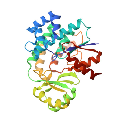
    6APF, 6APG, 6APK, 6APM

  • PubMed Abstract: 

    The crystal structure of the trans-acyltransferase (AT) from the disorazole polyketide synthase (PKS) was determined at room temperature to a resolution of 2.5 Å using a new method for the direct delivery of the sample into an X-ray free-electron laser. A novel sample extractor efficiently delivered limited quantities of microcrystals directly from the native crystallization solution into the X-ray beam at room temperature. The AT structure revealed important catalytic features of this core PKS enzyme, including the occurrence of conformational changes around the active site. The implications of these conformational changes for polyketide synthase reaction dynamics are discussed.


  • Organizational Affiliation

    Stanford Synchrotron Radiation Lightsource , 2575 Sand Hill Road, Menlo Park, California 94025, United States.


Macromolecules
Find similar proteins by:  (by identity cutoff)  |  3D Structure
Entity ID: 1
MoleculeChains Sequence LengthOrganismDetailsImage
DisD protein282Sorangium cellulosumMutation(s): 0 
Gene Names: dszDdisD
EC: 2.3.1.39
UniProt
Find proteins for Q4U443 (Sorangium cellulosum)
Explore Q4U443 
Go to UniProtKB:  Q4U443
Entity Groups  
Sequence Clusters30% Identity50% Identity70% Identity90% Identity95% Identity100% Identity
UniProt GroupQ4U443
Sequence Annotations
Expand
  • Reference Sequence
Experimental Data & Validation

Experimental Data

  • Method: X-RAY DIFFRACTION
  • Resolution: 2.50 Å
  • R-Value Free: 0.269 
  • R-Value Work: 0.225 
  • R-Value Observed: 0.230 
  • Space Group: P 21 21 21
Unit Cell:
Length ( Å )Angle ( ˚ )
a = 44.214α = 90
b = 54.642β = 90
c = 124.152γ = 90
Software Package:
Software NamePurpose
PHENIXrefinement
PHASERphasing
PDB_EXTRACTdata extraction
cctbx.xfeldata reduction
cctbx.primedata scaling
Cootmodel building

Structure Validation

View Full Validation Report


Entry History & Funding Information

Deposition Data


Funding OrganizationLocationGrant Number
National Institutes of Health/National Institute of General Medical Sciences (NIH/NIGMS)United StatesP41GM103393
Department of Energy (DOE, United States)United StatesDE-AC02-76SF00515

Revision History  (Full details and data files)

  • Version 1.0: 2018-04-04
    Type: Initial release
  • Version 2.0: 2018-04-11
    Changes: Data collection, Polymer sequence
  • Version 2.1: 2018-11-28
    Changes: Data collection
  • Version 2.2: 2019-12-04
    Changes: Author supporting evidence
  • Version 2.3: 2023-10-04
    Changes: Data collection, Database references, Refinement description