6L26

Neutron crystal structure of the mutant green fluorescent protein (EGFP)


Experimental Data Snapshot

  • Method: NEUTRON DIFFRACTION
  • Resolution: 1.44 Å
  • R-Value Free: 0.206 
  • R-Value Work: 0.168 
  • R-Value Observed: 0.170 

wwPDB Validation   3D Report Full Report


This is version 2.1 of the entry. See complete history


Literature

Direct Observation of the Protonation States in the Mutant Green Fluorescent Protein.

Shibazaki, C.Shimizu, R.Kagotani, Y.Ostermann, A.Schrader, T.E.Adachi, M.

(2020) J Phys Chem Lett 11: 492-496

  • DOI: https://doi.org/10.1021/acs.jpclett.9b03252
  • Primary Citation of Related Structures:  
    6L26, 6L27

  • PubMed Abstract: 

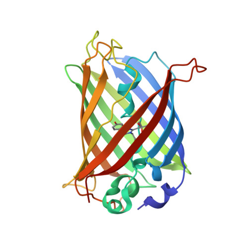
    Neutron crystallography has been used to elucidate the protonation states for the enhanced green fluorescent protein, which has revolutionized imaging technologies. The structure has a deprotonated hydroxyl group in the fluorescent chromophore. Also, the protonation states of His148 and Thr203, as well as the orientation of a critical water molecule in direct contact with the chromophore, could be determined. The results demonstrate that the deprotonated hydroxyl group in the chromophore and the nitrogen atom ND1 in His148 are charged negatively and positively, respectively, forming an ion pair. The position of the two deuterium atoms in the critical water molecule appears to be displaced slightly toward the acceptor oxygen atoms according to their omit maps. This displacement implies the formation of an intriguing electrostatic potential realized inside of the protein. Our findings provide new insights into future protein design strategies along with developments in quantum chemical calculations.


  • Organizational Affiliation

    Institute for Quantum Life Science , National Institutes for Quantum and Radiological Science and Technology (QST) , 2-4 Shirakata , Tokai , Ibaraki 319-1106 , Japan.


Macromolecules
Find similar proteins by:  (by identity cutoff)  |  3D Structure
Entity ID: 1
MoleculeChains Sequence LengthOrganismDetailsImage
Green fluorescent protein227Nicotiana benthamianaMutation(s): 1 
Gene Names: gfp
UniProt
Find proteins for W6KDG8 (Nicotiana benthamiana)
Explore W6KDG8 
Go to UniProtKB:  W6KDG8
Entity Groups  
Sequence Clusters30% Identity50% Identity70% Identity90% Identity95% Identity100% Identity
UniProt GroupW6KDG8
Sequence Annotations
Expand
  • Reference Sequence
Small Molecules
Ligands 1 Unique
IDChains Name / Formula / InChI Key2D Diagram3D Interactions
D3O (Subject of Investigation/LOI)
Query on D3O

Download Ideal Coordinates CCD File 
B [auth A]trideuteriooxidanium
D3 O
XLYOFNOQVPJJNP-ZRLBSURWSA-O
Modified Residues  1 Unique
IDChains TypeFormula2D DiagramParent
CRO
Query on CRO
A
L-PEPTIDE LINKINGC15 H17 N3 O5THR, TYR, GLY
Experimental Data & Validation

Experimental Data

  • Method: NEUTRON DIFFRACTION
  • Resolution: 1.44 Å
  • R-Value Free: 0.206 
  • R-Value Work: 0.168 
  • R-Value Observed: 0.170 
  • Space Group: P 21 21 21
Unit Cell:
Length ( Å )Angle ( ˚ )
a = 50.857α = 90
b = 62.16β = 90
c = 69.202γ = 90
Software Package:
Software NamePurpose
PHENIXrefinement
HKL-2000data reduction
SCALEPACKdata scaling
PHENIXphasing

Structure Validation

View Full Validation Report


Entry History & Funding Information

Deposition Data


Funding OrganizationLocationGrant Number
Ministry of Education, Culture, Sports, Science and Technology (Japan)JapanJP16KT0063

Revision History  (Full details and data files)

  • Version 1.0: 2020-04-08
    Type: Initial release
  • Version 2.0: 2021-06-09
    Changes: Non-polymer description
  • Version 2.1: 2023-11-22
    Changes: Data collection, Database references, Refinement description