6LON

Crystal structure of HPSG


Experimental Data Snapshot

  • Method: X-RAY DIFFRACTION
  • Resolution: 2.20 Å
  • R-Value Free: 0.179 
  • R-Value Work: 0.143 
  • R-Value Observed: 0.143 

wwPDB Validation   3D Report Full Report


Ligand Structure Quality Assessment 


This is version 1.3 of the entry. See complete history


Literature

Two radical-dependent mechanisms for anaerobic degradation of the globally abundant organosulfur compound dihydroxypropanesulfonate.

Liu, J.Wei, Y.Lin, L.Teng, L.Yin, J.Lu, Q.Chen, J.Zheng, Y.Li, Y.Xu, R.Zhai, W.Liu, Y.Liu, Y.Cao, P.Ang, E.L.Zhao, H.Yuchi, Z.Zhang, Y.

(2020) Proc Natl Acad Sci U S A 117: 15599-15608

  • DOI: https://doi.org/10.1073/pnas.2003434117
  • Primary Citation of Related Structures:  
    6LON

  • PubMed Abstract: 

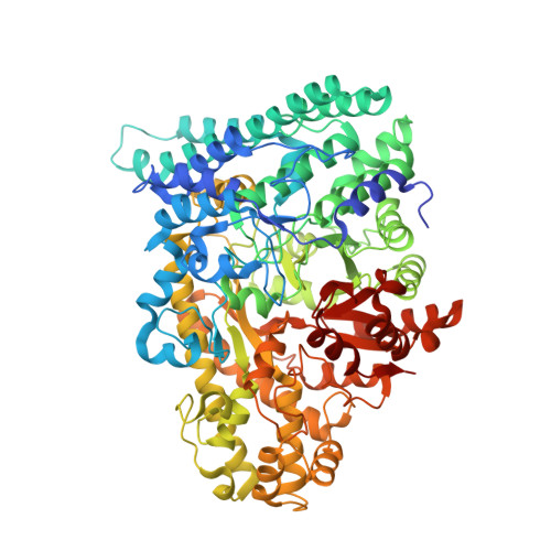
    2( S )-dihydroxypropanesulfonate (DHPS) is a microbial degradation product of 6-deoxy-6-sulfo-d-glucopyranose (sulfoquinovose), a component of plant sulfolipid with an estimated annual production of 10 10 tons. DHPS is also at millimolar levels in highly abundant marine phytoplankton. Its degradation and sulfur recycling by microbes, thus, play important roles in the biogeochemical sulfur cycle. However, DHPS degradative pathways in the anaerobic biosphere are not well understood. Here, we report the discovery and characterization of two O 2 -sensitive glycyl radical enzymes that use distinct mechanisms for DHPS degradation. DHPS-sulfolyase (HpsG) in sulfate- and sulfite-reducing bacteria catalyzes C-S cleavage to release sulfite for use as a terminal electron acceptor in respiration, producing H 2 S. DHPS-dehydratase (HpfG), in fermenting bacteria, catalyzes C-O cleavage to generate 3-sulfopropionaldehyde, subsequently reduced by the NADH-dependent sulfopropionaldehyde reductase (HpfD). Both enzymes are present in bacteria from diverse environments including human gut, suggesting the contribution of enzymatic radical chemistry to sulfur flux in various anaerobic niches.


  • Organizational Affiliation

    Tianjin Key Laboratory for Modern Drug Delivery & High-Efficiency, Collaborative Innovation Center of Chemical Science and Engineering, School of Pharmaceutical Science and Technology, Tianjin University, 300072 Tianjin, China.


Macromolecules
Find similar proteins by:  (by identity cutoff)  |  3D Structure
Entity ID: 1
MoleculeChains Sequence LengthOrganismDetailsImage
PFL2/glycerol dehydratase family glycyl radical enzymeA [auth B],
B [auth A],
C,
D
824Bilophila wadsworthia 3_1_6Mutation(s): 4 
Gene Names: HMPREF0179_02149
UniProt
Find proteins for E5Y7I4 (Bilophila wadsworthia (strain 3_1_6))
Explore E5Y7I4 
Go to UniProtKB:  E5Y7I4
Entity Groups  
Sequence Clusters30% Identity50% Identity70% Identity90% Identity95% Identity100% Identity
UniProt GroupE5Y7I4
Sequence Annotations
Expand
  • Reference Sequence
Small Molecules
Ligands 3 Unique
IDChains Name / Formula / InChI Key2D Diagram3D Interactions
LLQ (Subject of Investigation/LOI)
Query on LLQ

Download Ideal Coordinates CCD File 
AB [auth D],
E [auth B],
OA [auth C],
V [auth A]
(2~{S})-2,3-bis(oxidanyl)propane-1-sulfonic acid
C3 H8 O5 S
YPFUJZAAZJXMIP-VKHMYHEASA-N
GOL
Query on GOL

Download Ideal Coordinates CCD File 
AA [auth A]
BA [auth A]
BB [auth D]
CA [auth A]
CB [auth D]
AA [auth A],
BA [auth A],
BB [auth D],
CA [auth A],
CB [auth D],
DA [auth A],
DB [auth D],
EA [auth A],
EB [auth D],
F [auth B],
FA [auth A],
FB [auth D],
G [auth B],
GA [auth A],
GB [auth D],
H [auth B],
HA [auth A],
HB [auth D],
I [auth B],
IA [auth A],
IB [auth D],
J [auth B],
JA [auth A],
JB [auth D],
K [auth B],
KB [auth D],
L [auth B],
LA [auth A],
M [auth B],
MA [auth A],
MB [auth D],
N [auth B],
NA [auth A],
NB [auth D],
O [auth B],
P [auth B],
PA [auth C],
Q [auth B],
QA [auth C],
R [auth B],
RA [auth C],
S [auth B],
SA [auth C],
TA [auth C],
U [auth B],
UA [auth C],
VA [auth C],
W [auth A],
WA [auth C],
X [auth A],
XA [auth C],
Y [auth A],
YA [auth C],
Z [auth A]
GLYCEROL
C3 H8 O3
PEDCQBHIVMGVHV-UHFFFAOYSA-N
NA
Query on NA

Download Ideal Coordinates CCD File 
KA [auth A],
LB [auth D],
T [auth B],
ZA [auth C]
SODIUM ION
Na
FKNQFGJONOIPTF-UHFFFAOYSA-N
Binding Affinity Annotations 
IDSourceBinding Affinity
LLQ Binding MOAD:  6LON Kd: 6700 (nM) from 1 assay(s)
Experimental Data & Validation

Experimental Data

  • Method: X-RAY DIFFRACTION
  • Resolution: 2.20 Å
  • R-Value Free: 0.179 
  • R-Value Work: 0.143 
  • R-Value Observed: 0.143 
  • Space Group: C 1 2 1
Unit Cell:
Length ( Å )Angle ( ˚ )
a = 92.917α = 90
b = 219.401β = 98.04
c = 213.141γ = 90
Software Package:
Software NamePurpose
PHENIXrefinement
HKL-2000data scaling
PDB_EXTRACTdata extraction
HKL-3000data reduction
PHENIXphasing

Structure Validation

View Full Validation Report


Ligand Structure Quality Assessment 


Entry History & Funding Information

Deposition Data


Funding OrganizationLocationGrant Number
National Science Foundation (NSF, China)China31870049

Revision History  (Full details and data files)

  • Version 1.0: 2020-06-24
    Type: Initial release
  • Version 1.1: 2020-07-08
    Changes: Database references
  • Version 1.2: 2020-07-22
    Changes: Database references
  • Version 1.3: 2023-11-29
    Changes: Data collection, Database references, Derived calculations, Refinement description