6PV6

Functional Pathways of Biomolecules Retrieved from Single-particle Snapshots


Experimental Data Snapshot

  • Method: ELECTRON MICROSCOPY
  • Resolution: 4.50 Å
  • Aggregation State: 3D ARRAY 
  • Reconstruction Method: SINGLE PARTICLE 

wwPDB Validation   3D Report Full Report


This is version 1.2 of the entry. See complete history


Literature

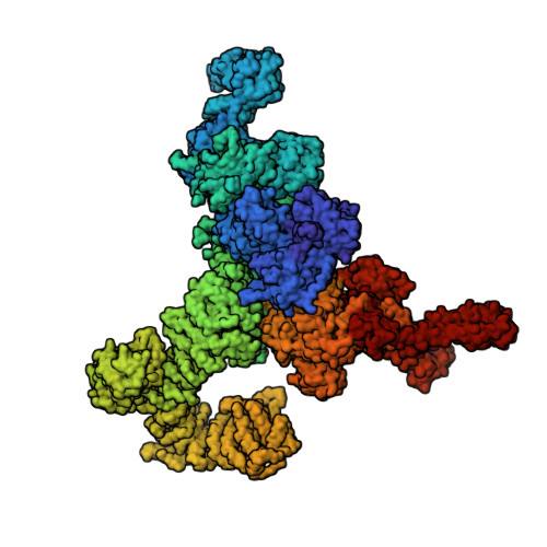
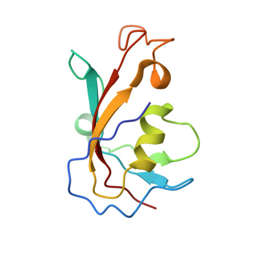
Retrieving functional pathways of biomolecules from single-particle snapshots.

Dashti, A.Mashayekhi, G.Shekhar, M.Ben Hail, D.Salah, S.Schwander, P.des Georges, A.Singharoy, A.Frank, J.Ourmazd, A.

(2020) Nat Commun 11: 4734-4734

  • DOI: https://doi.org/10.1038/s41467-020-18403-x
  • Primary Citation of Related Structures:  
    6PV6, 7JMF, 7JMG, 7JMH, 7JMI, 7JMJ

  • PubMed Abstract: 

    A primary reason for the intense interest in structural biology is the fact that knowledge of structure can elucidate macromolecular functions in living organisms. Sustained effort has resulted in an impressive arsenal of tools for determining the static structures. But under physiological conditions, macromolecules undergo continuous conformational changes, a subset of which are functionally important. Techniques for capturing the continuous conformational changes underlying function are essential for further progress. Here, we present chemically-detailed conformational movies of biological function, extracted data-analytically from experimental single-particle cryo-electron microscopy (cryo-EM) snapshots of ryanodine receptor type 1 (RyR1), a calcium-activated calcium channel engaged in the binding of ligands. The functional motions differ substantially from those inferred from static structures in the nature of conformationally active structural domains, the sequence and extent of conformational motions, and the way allosteric signals are transduced within and between domains. Our approach highlights the importance of combining experiment, advanced data analysis, and molecular simulations.


  • Organizational Affiliation

    Department of Physics, University of Wisconsin Milwaukee, 3135 N. Maryland Ave, Milwaukee, WI, 53211, USA.


Macromolecules
Find similar proteins by:  (by identity cutoff)  |  3D Structure
Entity ID: 1
MoleculeChains Sequence LengthOrganismDetailsImage
Ryanodine receptor 1A [auth B],
B [auth E],
C [auth I],
D [auth G]
4,687Oryctolagus cuniculusMutation(s): 0 
Entity Groups  
Sequence Clusters30% Identity50% Identity70% Identity90% Identity95% Identity100% Identity
Sequence Annotations
Expand
  • Reference Sequence
Find similar proteins by:  (by identity cutoff)  |  3D Structure
Entity ID: 2
MoleculeChains Sequence LengthOrganismDetailsImage
Peptidyl-prolyl cis-trans isomerase FKBP1BE [auth F],
F [auth A],
G [auth H],
H [auth J]
108Homo sapiensMutation(s): 0 
Gene Names: FKBP1BFKBP12.6FKBP1LFKBP9OTK4
EC: 5.2.1.8
UniProt & NIH Common Fund Data Resources
Find proteins for P68106 (Homo sapiens)
Explore P68106 
Go to UniProtKB:  P68106
PHAROS:  P68106
GTEx:  ENSG00000119782 
Entity Groups  
Sequence Clusters30% Identity50% Identity70% Identity90% Identity95% Identity100% Identity
UniProt GroupP68106
Sequence Annotations
Expand
  • Reference Sequence
Experimental Data & Validation

Experimental Data

  • Method: ELECTRON MICROSCOPY
  • Resolution: 4.50 Å
  • Aggregation State: 3D ARRAY 
  • Reconstruction Method: SINGLE PARTICLE 
EM Software:
TaskSoftware PackageVersion
MODEL REFINEMENTPHENIX

Structure Validation

View Full Validation Report


Entry History 

Deposition Data

Revision History  (Full details and data files)

  • Version 1.0: 2020-08-12
    Type: Initial release
  • Version 1.1: 2020-10-07
    Changes: Database references
  • Version 1.2: 2024-03-20
    Changes: Data collection, Database references, Refinement description