High-resolution crystal structures of a "half sandwich"-type Ru(II) coordination compound bound to hen egg-white lysozyme and proteinase K.
Chiniadis, L., Bratsos, I., Bethanis, K., Karpusas, M., Giastas, P., Papakyriakou, A.(2020) J Biol Inorg Chem 25: 635-645
- PubMed: 32266561
- DOI: https://doi.org/10.1007/s00775-020-01786-z
- Primary Citation of Related Structures:
6TVL, 6TXG - PubMed Abstract:
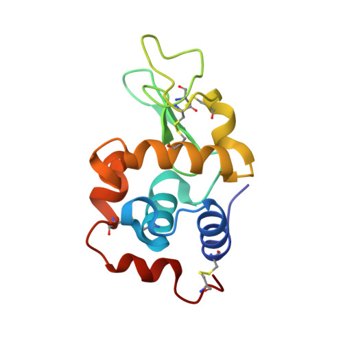
The high-resolution X-ray crystal structures of the adducts formed between the "half sandwich"-type Ru(II) coordination compound [Ru II (1,4,7-trithiacyclononane)(ethane-1,2-diamine)Cl] + and two proteins, namely hen egg-white lysozyme and proteinase K, are presented. The structures unveil that upon reaction with both enzymes the Ru(II) compound is coordinated by solvent-exposed aspartate residues after releasing the chloride ligand (Asp101 in lysozyme, Asp200 and Asp260 in proteinase K), while retaining the two chelating ligands. The adduct with Asp101 residue at the catalytic cleft of lysozyme is accompanied by residue-specific conformational changes to accommodate the Ru(II) fragment, whereas the complexes bound at the two calcium-binding sites of proteinase K revealed minimal structural perturbation of the enzyme. To the best of our knowledge, proteinase K is used here for the first time as a model system of protein metalation and these are the first X-ray crystal structures of protein adducts of a Ru(II) coordination compound that maintains its coordination sphere almost intact upon binding. Our data demonstrate the role of ligands in stabilizing the protein adducts via hydrophobic/aromatic or hydrogen-bonding interactions, as well as their underlying role in the selection of specific sites on the electrostatic potential surface of the enzymes.
Organizational Affiliation:
Institute of Biosciences and Applications, National Centre for Scientific Research "Demokritos", 15341, Athens, Greece.