6W6O

NaChBac-Nav1.7VSDII chimera and HWTX-IV complex


Experimental Data Snapshot

  • Method: ELECTRON MICROSCOPY
  • Resolution: 3.20 Å
  • Aggregation State: PARTICLE 
  • Reconstruction Method: SINGLE PARTICLE 

wwPDB Validation   3D Report Full Report


This is version 1.1 of the entry. See complete history


Literature

Employing NaChBac for cryo-EM analysis of toxin action on voltage-gated Na + channels in nanodisc.

Gao, S.Valinsky, W.C.On, N.C.Houlihan, P.R.Qu, Q.Liu, L.Pan, X.Clapham, D.E.Yan, N.

(2020) Proc Natl Acad Sci U S A 117: 14187-14193

  • DOI: https://doi.org/10.1073/pnas.1922903117
  • Primary Citation of Related Structures:  
    6VWX, 6VX3, 6VXO, 6W6O

  • PubMed Abstract: 

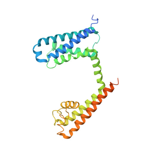
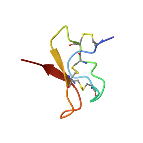
    NaChBac, the first bacterial voltage-gated Na + (Na v ) channel to be characterized, has been the prokaryotic prototype for studying the structure-function relationship of Na v channels. Discovered nearly two decades ago, the structure of NaChBac has not been determined. Here we present the single particle electron cryomicroscopy (cryo-EM) analysis of NaChBac in both detergent micelles and nanodiscs. Under both conditions, the conformation of NaChBac is nearly identical to that of the potentially inactivated Na v Ab. Determining the structure of NaChBac in nanodiscs enabled us to examine gating modifier toxins (GMTs) of Na v channels in lipid bilayers. To study GMTs in mammalian Na v channels, we generated a chimera in which the extracellular fragment of the S3 and S4 segments in the second voltage-sensing domain from Na v 1.7 replaced the corresponding sequence in NaChBac. Cryo-EM structures of the nanodisc-embedded chimera alone and in complex with HuwenToxin IV (HWTX-IV) were determined to 3.5 and 3.2 Å resolutions, respectively. Compared to the structure of HWTX-IV-bound human Na v 1.7, which was obtained at an overall resolution of 3.2 Å, the local resolution of the toxin has been improved from ∼6 to ∼4 Å. This resolution enabled visualization of toxin docking. NaChBac can thus serve as a convenient surrogate for structural studies of the interactions between GMTs and Na v channels in a membrane environment.


  • Organizational Affiliation

    Department of Molecular Biology, Princeton University, Princeton, NJ 08544.


Macromolecules
Find similar proteins by:  (by identity cutoff)  |  3D Structure
Entity ID: 1
MoleculeChains Sequence LengthOrganismDetailsImage
NaChBac-Nav1.7VSDII chimeraA,
C [auth D],
E [auth F],
G [auth H]
277Halalkalibacterium halodurans C-125Homo sapiensMutation(s): 0 
Gene Names: BH1501SCN9ANENA
Membrane Entity: Yes 
UniProt & NIH Common Fund Data Resources
Find proteins for Q15858 (Homo sapiens)
Explore Q15858 
Go to UniProtKB:  Q15858
PHAROS:  Q15858
GTEx:  ENSG00000169432 
Find proteins for Q9KCR8 (Halalkalibacterium halodurans (strain ATCC BAA-125 / DSM 18197 / FERM 7344 / JCM 9153 / C-125))
Explore Q9KCR8 
Go to UniProtKB:  Q9KCR8
Entity Groups  
Sequence Clusters30% Identity50% Identity70% Identity90% Identity95% Identity100% Identity
UniProt GroupsQ15858Q9KCR8
Sequence Annotations
Expand
  • Reference Sequence
Find similar proteins by:  (by identity cutoff)  |  3D Structure
Entity ID: 2
MoleculeChains Sequence LengthOrganismDetailsImage
Huwentoxin-IVB [auth C],
D [auth E],
F [auth G],
H [auth I]
35Cyriopagopus schmidtiMutation(s): 0 
Membrane Entity: Yes 
UniProt
Find proteins for P83303 (Cyriopagopus schmidti)
Explore P83303 
Go to UniProtKB:  P83303
Entity Groups  
Sequence Clusters30% Identity50% Identity70% Identity90% Identity95% Identity100% Identity
UniProt GroupP83303
Sequence Annotations
Expand
  • Reference Sequence
Small Molecules
Ligands 1 Unique
IDChains Name / Formula / InChI Key2D Diagram3D Interactions
POV
Query on POV

Download Ideal Coordinates CCD File 
I [auth A]
J [auth A]
K [auth A]
L [auth D]
M [auth D]
I [auth A],
J [auth A],
K [auth A],
L [auth D],
M [auth D],
N [auth D],
O [auth F],
P [auth F],
Q [auth F],
R [auth H],
S [auth H],
T [auth H]
(2S)-3-(hexadecanoyloxy)-2-[(9Z)-octadec-9-enoyloxy]propyl 2-(trimethylammonio)ethyl phosphate
C42 H82 N O8 P
WTJKGGKOPKCXLL-PFDVCBLKSA-N
Experimental Data & Validation

Experimental Data

  • Method: ELECTRON MICROSCOPY
  • Resolution: 3.20 Å
  • Aggregation State: PARTICLE 
  • Reconstruction Method: SINGLE PARTICLE 

Structure Validation

View Full Validation Report


Entry History & Funding Information

Deposition Data


Funding OrganizationLocationGrant Number
National Science Foundation (NSF, United States)United StatesDMR-1420541

Revision History  (Full details and data files)

  • Version 1.0: 2020-06-24
    Type: Initial release
  • Version 1.1: 2021-01-13
    Changes: Database references