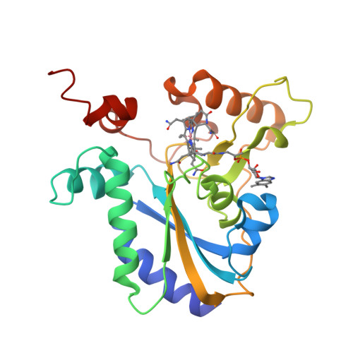
Warning You are using a web browser that we do not support. Our website will not work properly. Please update to a newer version or download a new web browser, such as Chrome or Firefox.
Help
Contact us
  • Sign in with Google
  • Sign in with Facebook
  • Sign in with ORCID

  • Help
RCSB PDB
  • Deposit 

      Prepare Data

    • PDBx/mmCIF file
    • pdb_extract
    • SF-Tool
    • Ligand Expo
    • MAXIT

      Validate Data

    • Validation Server
    • Validation API
    • Information for Journals
    • Validation Task Forces

      Deposit Data

    • wwPDB OneDep System
    • PDB-IHM

      Help and Resources

    • Deposit FAQ
    • Validation FAQ
    • Tutorials
    • Annotation Policies
    • Processing Procedures
    • PDBx/mmCIF Dictionary
    • PDBx/mmCIF User Guide
    • Chemical Component Dictionary
    • Biologically Interesting Molecule Reference Dictionary (BIRD)
    • BioSync/Beamlines/Facilities
    • Related Tools
  • Search 
    • Advanced Search
    • Sequence Similarity Search
    • Structure Similarity Search
    • Chemical Similarity Search
    • Chemical Sketch Tool
    • Browse by Annotations
    • New Entries
    • Unreleased Entries
    • PDB Statistics
  • Visualize 
    • Mol* (MolStar)
    • Sequence Annotations Viewer
    • Genome View
  • Analyze 
    • Pairwise Structure Alignment
    • Symmetry Resources in the PDB
    • Structure Quality
    • Grouping Structures
    • PDB Citation MeSH Network Explorer
    • PDB Statistics
    • EPPIC Biological Assemblies
    • External Data and Resources
    • Integrated Resources
    • Additional Resources
  • Download 
    • Coordinates and Experimental Data
    • Sequences
    • Ligands
    • File Download Services
    • Web APIs
  • Learn 

    • Training Courses
    • Guide to PDB Data
    • Molecule of the Month
    • Educational Resources
    • Curricula
    • Browse
    • News
    • SciArt Galleries
    • Irving Geis
    • David Goodsell
  • About 
    • Contact Us
    • About RCSB PDB
    • Vision and Mission
    • Citation, Usage, Privacy Policies, Logo
    • News
    • PDB History
    • PDB50
    • User Community
    • Publications
    • RCSB PDB Advisory Committee
    • Team Members
    • Service Status
  • Careers
  • COVID-19
RCSB PDB
239,865
Structures from the PDB archive
1,068,577
Computed Structure Models (CSM)
Include CSM
Advanced Search
|
Browse Annotations
Help
  • PDB-101
  • wwPDB
  • EMDataResource
  • NAKB: Nucleic Acid Knowledgebase
  • wwPDB Foundation
  • PDB-IHM Logo
  • Search
  • Query History
  • Browse Annotations
  • MyPDB
QUERY: Structure Author = "Banerjee, R."
MyPDB Login
 Search API
Advanced Search Query Builder
Help
Full Text
Structure Attributes
Help
x
NOT
Count
Add Subquery
Chemical Attributes
Sequence Similarity
Sequence Motif
Structure Similarity
Structure Motif
Chemical Similarity
Return
grouped by
Include Computed Structure Models (CSM)
Count
Clear
Search
Search Summary This query matches 124 Structures.
Refinements
Structure Determination Methodology
Scientific Name of Source Organism
More...
Taxonomy
Experimental Method
Polymer Entity Type
Refinement Resolution (Å)
Release Date
Enzyme Classification Name
Membrane Protein Annotation
Symmetry Type
SCOP Classification
1 to 25 of 124 Structures
Page 1 of 5
Sort by
Gallery item
 Explore in 3D

1JAC | pdb_00001jac

Download FileView File

A NOVEL MODE OF CARBOHYDRATE RECOGNITION IN JACALIN, A MORACEAE PLANT LECTIN WITH A BETA-PRISM

Sankaranarayanan, R., Sekar, K., Banerjee, R., Sharma, V., Surolia, A., Vijayan, M.

(1996) Nat Struct Biol 3: 596-603

Released1997-06-05
Method
X-RAY DIFFRACTION 2.43 Å
Organisms
Artocarpus heterophyllus
Macromolecule
JACALIN (protein)
Unique LigandsAMG
Gallery item
 Explore in 3D

1M54 | pdb_00001m54

Download FileView File

CYSTATHIONINE-BETA SYNTHASE: REDUCED VICINAL THIOLS

Taoka, S., Lepore, B.W., Kabil, O., Ojha, S., Ringe, D., Banerjee, R.

(2002) Biochemistry 41: 10454-10461

Released2002-08-14
Method
X-RAY DIFFRACTION 2.9 Å
Organisms
Homo sapiens
Macromolecule
CYSTATHIONINE BETA-SYNTHASE (protein)
Unique LigandsHEM, PLP
Gallery item
 Explore in 3D

2HAQ | pdb_00002haq

Download FileView File

Crystal Structure of Cyclophilin A from Leishmania Donovani

Venugopal, V., Sen, B., Datta, A.K., Banerjee, R.

(2007) Acta Crystallogr Sect F Struct Biol Cryst Commun 63: 60-64

Released2006-06-20
Method
X-RAY DIFFRACTION 1.97 Å
Organisms
Leishmania donovani
Macromolecule
Cyclophilin (protein)
Gallery item
 Explore in 3D

2M6U | pdb_00002m6u

Download FileView File

NMR Structure of CbpAN from Streptococcus pneumoniae

Liu, A., Yan, H., Achila, D., Martinez-Hackert, E., Li, Y., Banerjee, R.

(2015) Biochem J 465: 325-335

Released2014-04-16
Method
SOLUTION NMR
Organisms
Streptococcus pneumoniae
Macromolecule
Choline binding protein A (protein)
Gallery item
 Explore in 3D

2PEL | pdb_00002pel

Download FileView File

PEANUT LECTIN

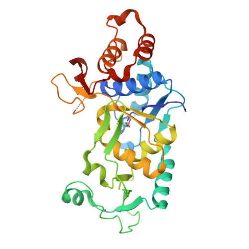
Banerjee, R., Das, K., Ravishankar, R., Suguna, K., Surolia, A., Vijayan, M.

(1996) J Mol Biology 259: 281-296

Released1996-12-07
Method
X-RAY DIFFRACTION 2.25 Å
Organisms
Arachis hypogaea
Macromolecule
PEANUT LECTIN (protein)
Unique LigandsCA, MN
Unique branched monosaccharidesBGC, GAL, GLC
Gallery item
 Explore in 3D

2QM7 | pdb_00002qm7

Download FileView File

MeaB, A Bacterial Homolog of MMAA, Bound to GDP

Hubbard, P.A., Padovani, D., Labunska, T., Mahlstedt, S.A., Banerjee, R., Drennan, C.L.

(2007) J Biological Chem 282: 31308-31316

Released2007-08-28
Method
X-RAY DIFFRACTION 1.85 Å
Organisms
Methylorubrum extorquens AM1
Macromolecule
GTPase/ATPase (protein)
Unique LigandsGDP, PO4
Gallery item
 Explore in 3D

2QM8 | pdb_00002qm8

Download FileView File

MeaB, A Bacterial Homolog of MMAA, in the Nucleotide Free Form

Hubbard, P.A., Padovani, D., Labunska, T., Mahlstedt, S.A., Banerjee, R., Drennan, C.L.

(2007) J Biological Chem 282: 31308-31316

Released2007-08-28
Method
X-RAY DIFFRACTION 1.7 Å
Organisms
Methylorubrum extorquens AM1
Macromolecule
GTPase/ATPase (protein)
Unique LigandsPO4
Gallery item
 Explore in 3D

3BT8 | pdb_00003bt8

Download FileView File

Crystal Structure of Mutant Cyclophilin (R147A) from Leishmania donovani

Venugopal, V., Sen, B., Datta, A.K., Banerjee, R.

To be published

Released2008-01-15
Method
X-RAY DIFFRACTION 2.7 Å
Organisms
Leishmania donovani
Macromolecule
Peptidyl-prolyl cis-trans isomerase (protein)
Gallery item
 Explore in 3D

3EOV | pdb_00003eov

Download FileView File

Crystal structure of cyclophilin from Leishmania donovani ligated with cyclosporin A

Venugopal, V., Dasgupta, D., Datta, A.K., Banerjee, R.

(2009) Acta Crystallogr D Biol Crystallogr 65: 1187

Released2008-11-11
Method
X-RAY DIFFRACTION 2.6 Å
Organisms
Leishmania donovani
Tolypocladium inflatum
Macromolecule
CYCLOSPORIN A (protein)
PEPTIDYL-PROLYL CIS-TRANS ISOMERASE (protein)
Gallery item
 Explore in 3D

3RX6 | pdb_00003rx6

Download FileView File

Crystal structure of Polarity Suppression protein from Enterobacteria phage P4

Banerjee, R., Nath, S., Khamrui, S., Sen, R., Sen, U.

(2012) J Biological Chem 287: 44667-44675

Released2012-07-25
Method
X-RAY DIFFRACTION 2.039 Å
Organisms
Enterobacteria phage P4
Macromolecule
Polarity suppression protein (protein)
Unique LigandsHG, IOD, TRS
Gallery item
 Explore in 3D

3SBY | pdb_00003sby

Download FileView File

Crystal Structure of SeMet-Substituted Apo-MMACHC (1-244), a human B12 processing enzyme

Koutmos, M., Gherasim, C., Smith, J.L., Banerjee, R.

(2011) J Biological Chem 286: 29780-29787

Released2011-06-22
Method
X-RAY DIFFRACTION 2.71 Å
Organisms
Homo sapiens
Macromolecule
Methylmalonic aciduria and homocystinuria type C protein (protein)
Gallery item
 Explore in 3D

3SBZ | pdb_00003sbz

Download FileView File

Crystal Structure of Apo-MMACHC (1-244), a human B12 processing enzyme

Koutmos, M., Gherasim, C., Smith, J.L., Banerjee, R.

(2011) J Biological Chem 286: 29780-29787

Released2011-06-22
Method
X-RAY DIFFRACTION 2 Å
Organisms
Homo sapiens
Macromolecule
Methylmalonic aciduria and homocystinuria type C protein (protein)
Unique LigandsGOL, MLI
Gallery item
 Explore in 3D

3SC0 | pdb_00003sc0

Download FileView File

Crystal Structure of MMACHC (1-238), a human B12 processing enzyme, complexed with MethylCobalamin

Koutmos, M., Gherasim, C., Smith, J.L., Banerjee, R.

(2011) J Biological Chem 286: 29780-29787

Released2011-06-22
Method
X-RAY DIFFRACTION 1.95 Å
Organisms
Homo sapiens
Macromolecule
Methylmalonic aciduria and homocystinuria type C protein (protein)
Unique LigandsCOB
Gallery item
 Explore in 3D

4A91 | pdb_00004a91

Download FileView File

Crystal structure of the glutamyl-queuosine tRNAAsp synthetase from E. coli complexed with L-glutamate

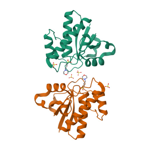
Blaise, M., Olieric, V., Sauter, C., Lorber, B., Roy, B., Karmakar, S., Banerjee, R., Becker, H.D., Kern, D.

(2008) J Mol Biology 381: 1224

Released2012-01-11
Method
X-RAY DIFFRACTION 1.75 Å
Organisms
Escherichia coli
Macromolecule
GLUTAMYL-Q TRNA(ASP) SYNTHETASE (protein)
Unique LigandsGLU, ZN
Gallery item
 Explore in 3D

4DVD | pdb_00004dvd

Download FileView File

Crystal structure of the disulphide linked knotted homodimer of Psu

Banerjee, R., Nath, S., Sen, U.

(2012) J Biological Chem 287: 44667-44675

Released2012-11-21
Method
X-RAY DIFFRACTION 3 Å
Organisms
Enterobacteria phage P4
Macromolecule
Polarity suppression protein (protein)
Gallery item
 Explore in 3D

4HI1 | pdb_00004hi1

Download FileView File

Crystal structure of acylphosphatase C20R mutant from Vibrio cholerae0395

Nath, S., Banerjee, R., Sen, U.

(2013) J Mol Biology

Released2013-09-25
Method
X-RAY DIFFRACTION 1.964 Å
Organisms
Vibrio cholerae O395
Macromolecule
Acylphosphatase (protein)
Unique LigandsMOO, SO4
Gallery item
 Explore in 3D

4HI2 | pdb_00004hi2

Download FileView File

Crystal structure of an Acylphosphatase protein cage

Nath, S., Banerjee, R., Sen, U.

(2013) J Mol Biology

Released2013-09-25
Method
X-RAY DIFFRACTION 3.1 Å
Organisms
Vibrio cholerae O395
Macromolecule
Acylphosphatase (protein)
Unique LigandsSO4
Gallery item
 Explore in 3D

4JGT | pdb_00004jgt

Download FileView File

Structure and kinetic analysis of H2S production by human Mercaptopyruvate Sulfurtransferase

Koutmos, M., Yamada, K., Yadav, P.K., Chiku, T., Banerjee, R.

(2013) J Biological Chem 288: 20002-20013

Released2013-05-29
Method
X-RAY DIFFRACTION 2.161 Å
Organisms
Homo sapiens
Macromolecule
3-mercaptopyruvate sulfurtransferase (protein)
Unique LigandsGOL, PYR, SO4
Gallery item
 Explore in 3D

4JYB | pdb_00004jyb

Download FileView File

MeaB, A Bacterial Homolog of MMAA, Bound to GMPPNP

Koutmos, M., Lofgren, M., Padovani, D., Banerjee, R.

(2013) Nat Chem Biol 9: 535-539

Released2013-07-24
Method
X-RAY DIFFRACTION 2.1 Å
Organisms
Methylorubrum extorquens AM1
Macromolecule
Methylmalonyl-CoA mutase accessory protein (protein)
Unique LigandsGNP
Gallery item
 Explore in 3D

4JYC | pdb_00004jyc

Download FileView File

MeaB, A Bacterial Homolog of MMAA, in its Apo form

Koutmos, M., Lofgren, M., Padovani, D., Banerjee, R.

(2013) Nat Chem Biol 9: 535-539

Released2013-07-24
Method
X-RAY DIFFRACTION 2.2 Å
Organisms
Methylorubrum extorquens AM1
Macromolecule
Methylmalonyl-CoA mutase accessory protein (protein)
Unique LigandsGDP
Gallery item
 Explore in 3D

4K12 | pdb_00004k12

Download FileView File

Structural Basis for Host Specificity of Factor H Binding by Streptococcus pneumoniae

Liu, A., Achila, D., Banerjee, R., Martinez-Hackert, E., Li, Y., Yan, H.

(2015) Biochem J 465: 325-335

Released2014-04-09
Method
X-RAY DIFFRACTION 1.079 Å
Organisms
Homo sapiens
Streptococcus pneumoniae
Macromolecule
Choline binding protein A (protein)
Complement factor H (protein)
Gallery item
 Explore in 3D

4LC1 | pdb_00004lc1

Download FileView File

MeaB, A Bacterial Homolog of MMAA, Bound to GDP and crystallized in the presence of GDP and [AlF4]-

Koutmos, M., Padovani, D., Lofgren, M., Banerjee, R.

(2013) J Biological Chem 288: 30980-30989

Released2013-09-25
Method
X-RAY DIFFRACTION 1.8 Å
Organisms
Methylorubrum extorquens AM1
Macromolecule
Methylmalonyl-CoA mutase accessory protein (protein)
Unique LigandsGDP, GOL
Gallery item
 Explore in 3D

4LRQ | pdb_00004lrq

Download FileView File

Crystal structure of a Low Molecular Weight Phosphotyrosine phosphatase from Vibrio choleraeO395

Nath, S., Banerjee, R., Sen, U.

(2014) Biochem Biophys Res Commun 450: 390-395

Released2014-07-02
Method
X-RAY DIFFRACTION 1.45 Å
Organisms
Vibrio cholerae O395
Macromolecule
Phosphotyrosine protein phosphatase (protein)
Unique LigandsMPO, SO4
Gallery item
 Explore in 3D

4QG5 | pdb_00004qg5

Download FileView File

Crystal structure of phosphoglucomutase from Leishmania major at 3.5 angstrom resolution

Waugh, B., Sen, U., Banerjee, R.

To be published

Released2015-05-27
Method
X-RAY DIFFRACTION 3.5 Å
Organisms
Leishmania major
Macromolecule
Putative phosphoglucomutase (protein)
Unique LigandsMG
Gallery item
 Explore in 3D

4S1E | pdb_00004s1e

Download FileView File

Crystal structure of cyclophilin mutant L120A from Leishmania donovani at 2.22 angstrom.

Roy, S., Datta, A.K., Banerjee, R.

To be published

Released2015-03-11
Method
X-RAY DIFFRACTION 2.22 Å
Organisms
Leishmania donovani
Macromolecule
Peptidyl-prolyl cis-trans isomerase (protein)
1 to 25 of 124 Structures
Page 1 of 5
Sort by
  • About
  • About Us
  • Citing Us
  • Publications
  • Team
  • Careers
  • Usage & Privacy
  • Support
  • Contact Us
  • Help
  • Website FAQ
  • Glossary
  • Service Status
  • RCSB PDB is hosted by

  • The Rutgers Artificial Intelligence and Data Science Collaboratory logo
    Uiversity of California San Diego logoSan Diego Supercomputer Center logo
    University of California San Francisco Logo
  • RCSB PDB is a member of the

  • wwPDB
    EMDataResource

  • RCSB Partners
  • Nucleic Acid Knowledgebase
  • wwPDB Partners
  • RCSB PDB
  • PDBe
  • PDBj
  • BMRB
  • EMDB
CoreTrustSeal
Global Biodata Coalition Logo

RCSB PDB Core Operations are funded by the U.S. National Science Foundation (DBI-2321666), the US Department of Energy (DE-SC0019749), and the National Cancer Institute, National Institute of Allergy and Infectious Diseases, and National Institute of General Medical Sciences of the National Institutes of Health under grant R01GM157729.