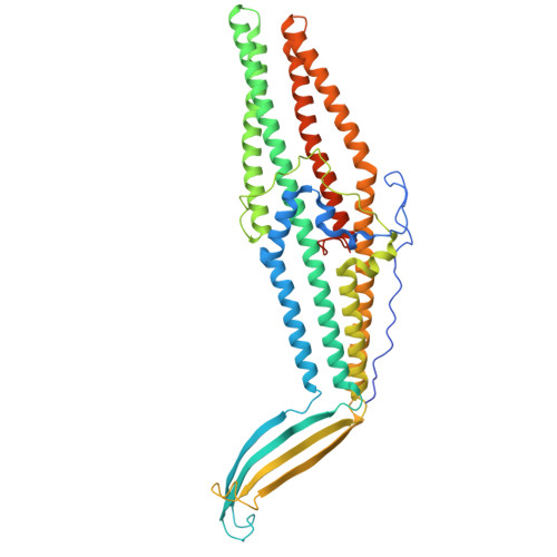
13GG | pdb_000013gg

High resolution cryo-EM structure of the Pseudomonas aeruginosa OprM Multidrug Efflux Pump


Experimental Data Snapshot

  • Method: ELECTRON MICROSCOPY
  • Resolution: 2.09 Å
  • Aggregation State: PARTICLE 
  • Reconstruction Method: SINGLE PARTICLE 

wwPDB Validation 3D Report Full Report

Validation slider image for 13GG

This is version 1.0 of the entry. See complete history. 

Literature

High resolution cryo-EM structure of the Pseudomonas aeruginosa OprM multidrug efflux pump provides target for rational inhibitor design.

Ratliff, A.C., Botos, I., Putti, A.M., Patil, P.P., Ramkumar, A., Ghirlando, R., Dekker, J.P., Buchanan, S.K.

(2026) J Struct Biol X 14: 100152-100152

  • DOI: https://doi.org/10.1016/j.yjsbx.2026.100152
  • Primary Citation Related Structures: 
    13GG

  • PubMed Abstract: 

    OprM is the outer membrane channel component of resistance-nodulation-division (RND) efflux pumps in Pseudomonas aeruginosa , a high-priority human pathogen. OprM-dependent pumps play diverse roles in bacterial physiology and mediate resistance to critical antibiotic classes, including fluoroquinolones, aminoglycosides, and broad-spectrum β-lactam/β-lactamase inhibitor combinations such as ceftazidime/avibactam. As the outer membrane component of the tripartite efflux system, OprM gates substrate export through an iris-like periplasmic pore that opens upon association with its inner membrane partners. Here we present a single-particle cryo-EM structure of OprM at 2.08 Å, the highest resolution OprM structure to date. The structure reveals a closed leucine-lined periplasmic pore, consistent with previous X-ray structures. A detailed structure-based comparison of the closed and open states reveals the conformational changes accompanying periplasmic pore opening, including iris-like twisting of the coiled-coil helices, displacement of the gating Leu429 residues, and a broader reorganization of salt bridges than previously described. Conservation of the MexA-binding interface across multiple Mex proteins suggests a shared mechanism of OprM engagement. Three lipopolysaccharide (LPS) molecules are resolved at the β-barrel interface, bridging adjacent OprM monomers through a combination of hydrophobic interactions with the fatty acyl chains and specific polar contacts with the KDO, heptose, and phosphate groups. This binding mode resembles that of other generic β-barrels and is expected to be preserved in vivo. Together, these findings provide the most detailed structural characterization of OprM to date, offering new insights into its gating mechanism and membrane interactions with implications for the development of efflux pump inhibitors.


  • Organizational Affiliation: 
    • Laboratory of Molecular Biology, National Institute of Diabetes & Digestive & Kidney Diseases (NIDDK), National Institutes of Health (NIH), Bethesda, MD 20892, USA.

Macromolecule Content 

  • Total Structure Weight: 172.62 kDa 
  • Atom Count: 11,073 
  • Modeled Residue Count: 1,368 
  • Deposited Residue Count: 1,500 
  • Unique protein chains: 1

Macromolecules

Find similar proteins by:|  3D Structure
Entity ID: 1
MoleculeChains  Sequence LengthOrganismDetailsImage
Outer membrane protein OprM
A, B, C
500Pseudomonas aeruginosa PAO1Mutation(s): 0 
Gene Names: oprM, oprK, PA0427
UniProt
Find proteins for Q51487 (Pseudomonas aeruginosa (strain ATCC 15692 / DSM 22644 / CIP 104116 / JCM 14847 / LMG 12228 / 1C / PRS 101 / PAO1))
Explore Q51487 
Go to UniProtKB:  Q51487
Entity Groups
Sequence Clusters30% Identity50% Identity70% Identity90% Identity95% Identity100% Identity
UniProt GroupQ51487
Sequence Annotations
Expand
Reference Sequence

Small Molecules

Ligands 1 Unique
IDChains Name / Formula / InChI Key2D Diagram3D Interactions
JSG

Query on JSG


Download:Ideal Coordinates CCD File
D [auth A],
E [auth A],
F [auth B]
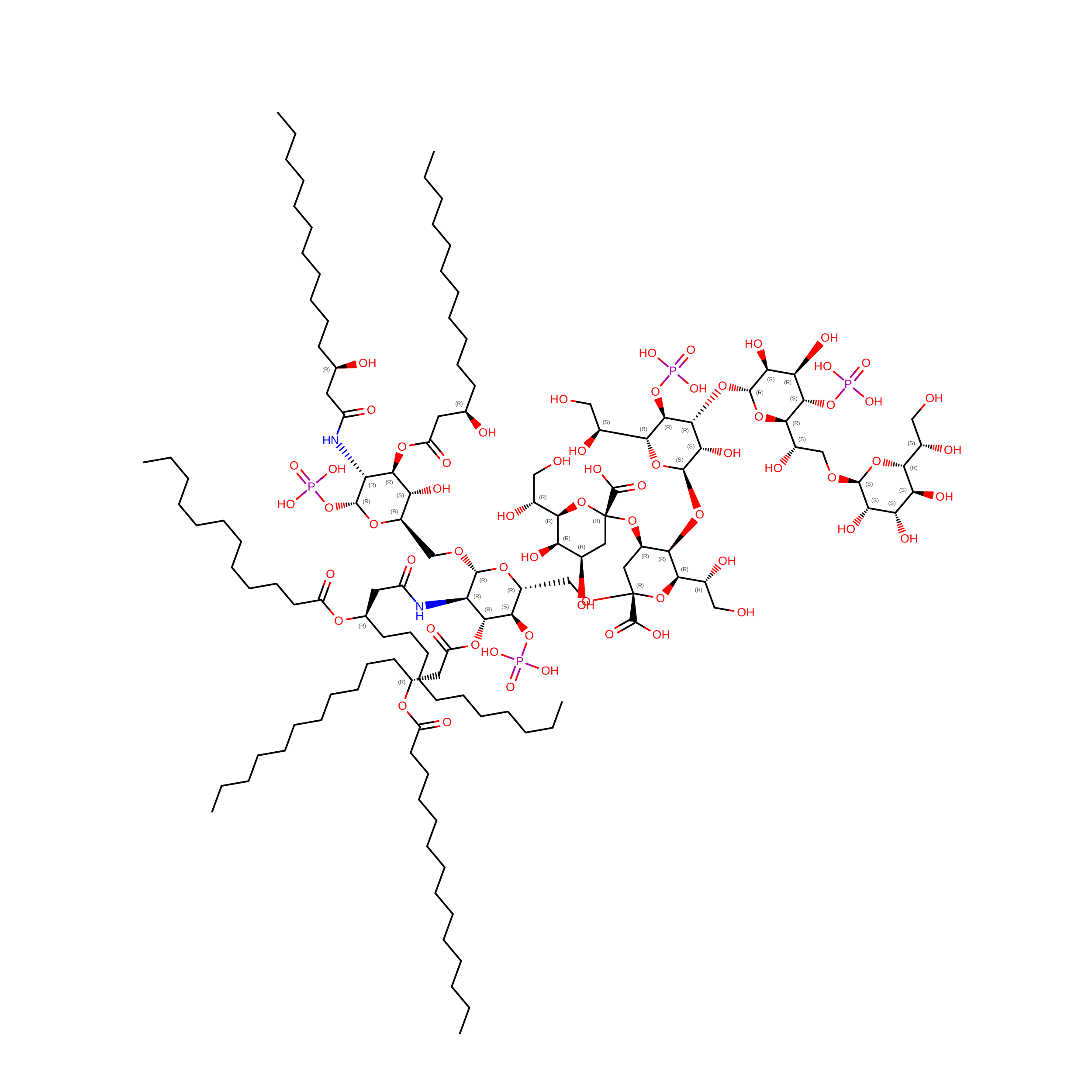
(2~{R},4~{R},5~{R},6~{R})-6-[(1~{R})-1,2-bis(oxidanyl)ethyl]-2-[(2~{R},4~{R},5~{R},6~{R})-6-[(1~{R})-1,2-bis(oxidanyl)ethyl]-5-[(2~{S},3~{S},4~{R},5~{R},6~{R})-6-[(1~{S})-1,2-bis(oxidanyl)ethyl]-4-[(2~{R},3~{S},4~{R},5~{S},6~{R})-6-[(1~{S})-2-[(2~{S},3~{S},4~{S},5~{S},6~{R})-6-[(1~{S})-1,2-bis(oxidanyl)ethyl]-3,4,5-tris(oxidanyl)oxan-2-yl]oxy-1-oxidanyl-ethyl]-3,4-bis(oxidanyl)-5-phosphonooxy-oxan-2-yl]oxy-3-oxidanyl-5-phosphonooxy-oxan-2-yl]oxy-2-carboxy-2-[[(2~{R},3~{S},4~{R},5~{R},6~{R})-5-[[(3~{R})-3-dodecanoyloxytetradecanoyl]amino]-6-[[(2~{R},3~{S},4~{R},5~{R},6~{R})-3-oxidanyl-5-[[(3~{R})-3-oxidanyltetradecanoyl]amino]-4-[(3~{R})-3-oxidanyltetradecanoyl]oxy-6-phosphonooxy-oxan-2-yl]methoxy]-3-phosphonooxy-4-[(3~{R})-3-tetradecanoyloxytetradecanoyl]oxy-oxan-2-yl]methoxy]oxan-4-yl]oxy-4,5-bis(oxidanyl)oxane-2-carboxylic acid
C131 H240 N2 O63 P4
MVYTXVAKEAWEGM-SMTLFHDCSA-N

Experimental Data & Validation

Experimental Data

  • Method: ELECTRON MICROSCOPY
  • Resolution: 2.09 Å
  • Aggregation State: PARTICLE 
  • Reconstruction Method: SINGLE PARTICLE 
EM Software:
TaskSoftware PackageVersion
RECONSTRUCTIONPHENIX2.21
RECONSTRUCTIONISOLDE
RECONSTRUCTIONCoot

Structure Validation

View Full Validation Report


Entry History 

& Funding Information

Deposition Data


Funding OrganizationLocationGrant Number
National Institutes of Health/National Institute of Diabetes and Digestive and Kidney Disease (NIH/NIDDK)United StatesZIA DK011011-17

Revision History  (Full details and data files)

  • Version 1.0: 2026-11-18
    Type: Initial release