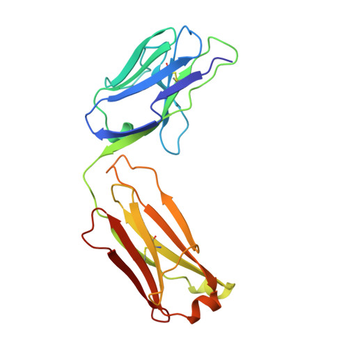
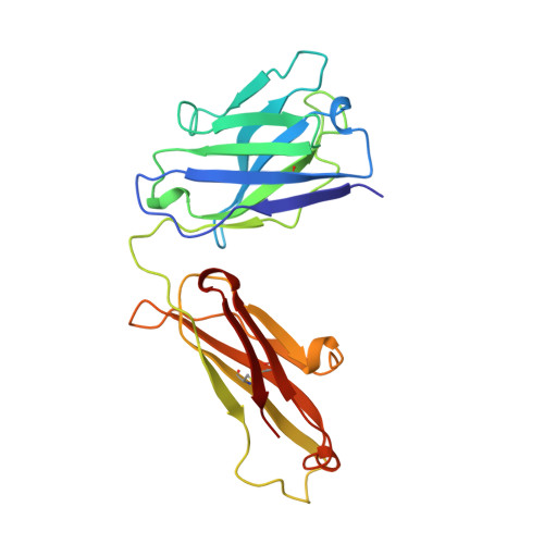
1AD0

FAB FRAGMENT OF ENGINEERED HUMAN MONOCLONAL ANTIBODY A5B7


Experimental Data Snapshot

  • Method: X-RAY DIFFRACTION
  • Resolution: 2.50 Å
  • R-Value Free: 0.262 
  • R-Value Work: 0.209 
  • R-Value Observed: 0.209 

Starting Model: experimental
View more details

wwPDB Validation   3D Report Full Report


This is version 1.4 of the entry. See complete history


Literature

VL:VH domain rotations in engineered antibodies: crystal structures of the Fab fragments from two murine antitumor antibodies and their engineered human constructs.

Banfield, M.J.King, D.J.Mountain, A.Brady, R.L.

(1997) Proteins 29: 161-171

  • DOI: https://doi.org/10.1002/(sici)1097-0134(199710)29:2<161::aid-prot4>3.0.co;2-g
  • Primary Citation of Related Structures:  
    1AD0, 1AD9, 1AE6, 1CLO

  • PubMed Abstract: 

    The crystal structures of two pairs of Fab fragments have been determined. The pairs comprise both a murine and an engineered human form, each derived from the antitumor antibodies A5B7 and CTM01. Although antigen specificity is maintained within the pairs, antigen affinity varies. A comparison of the hypervariable loops for each pair of antibodies shows their structure has been well maintained in grafting, supporting the canonical loop model. Detailed structural analysis of the binding sites and domain arrangements for these antibodies suggests the differences in antigen affinity observed are likely to be due to inherent flexibility of the hypervariable loops and movements at the VL:VH domain interface. The four structures provide the first opportunity to study in detail the effects of protein engineering on specific antibodies.


  • Organizational Affiliation

    Molecular Recognition Centre, University of Bristol School of Medical Sciences, United Kingdom.


Macromolecules
Find similar proteins by:  (by identity cutoff)  |  3D Structure
Entity ID: 1
MoleculeChains Sequence LengthOrganismDetailsImage
ANTIBODY A5B7 (LIGHT CHAIN)
A, C
213Homo sapiensMutation(s): 0 
Entity Groups  
Sequence Clusters30% Identity50% Identity70% Identity90% Identity95% Identity100% Identity
Sequence Annotations
Expand
  • Reference Sequence
Find similar proteins by:  (by identity cutoff)  |  3D Structure
Entity ID: 2
MoleculeChains Sequence LengthOrganismDetailsImage
ANTIBODY A5B7 (HEAVY CHAIN)
B, D
220Homo sapiensMutation(s): 0 
Entity Groups  
Sequence Clusters30% Identity50% Identity70% Identity90% Identity95% Identity100% Identity
Sequence Annotations
Expand
  • Reference Sequence
Experimental Data & Validation

Experimental Data

  • Method: X-RAY DIFFRACTION
  • Resolution: 2.50 Å
  • R-Value Free: 0.262 
  • R-Value Work: 0.209 
  • R-Value Observed: 0.209 
  • Space Group: P 1
Unit Cell:
Length ( Å )Angle ( ˚ )
a = 48.31α = 69.88
b = 66.27β = 82.48
c = 81.38γ = 71.34
Software Package:
Software NamePurpose
DENZOdata reduction
SCALEPACKdata scaling
X-PLORmodel building
X-PLORrefinement
X-PLORphasing

Structure Validation

View Full Validation Report


Entry History 

Deposition Data

Revision History  (Full details and data files)

  • Version 1.0: 1998-02-25
    Type: Initial release
  • Version 1.1: 2008-03-24
    Changes: Version format compliance
  • Version 1.2: 2011-07-13
    Changes: Version format compliance
  • Version 1.3: 2023-08-02
    Changes: Database references, Refinement description
  • Version 1.4: 2024-11-20
    Changes: Data collection, Structure summary