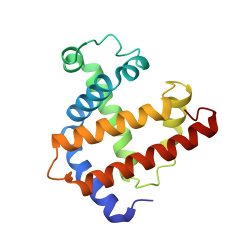
1AJG

CARBONMONOXY MYOGLOBIN AT 40 K


Experimental Data Snapshot

  • Method: X-RAY DIFFRACTION
  • Resolution: 1.69 Å
  • R-Value Free: 0.238 
  • R-Value Work: 0.171 
  • R-Value Observed: 0.171 

Starting Model: experimental
View more details

wwPDB Validation   3D Report Full Report


Ligand Structure Quality Assessment 


This is version 1.4 of the entry. See complete history


Literature

Photolysis-induced structural changes in single crystals of carbonmonoxy myoglobin at 40 K.

Teng, T.Y.Srajer, V.Moffat, K.

(1994) Nat Struct Biol 1: 701-705

  • DOI: https://doi.org/10.1038/nsb1094-701
  • Primary Citation of Related Structures:  
    1AJG, 1AJH

  • PubMed Abstract: 

    Myoglobin's reversible binding of oxygen is a model for studies of protein control of ligand binding and discrimination. Protein relaxation and geminate ligand rebinding subsequent to ligand photodissociation have been studied extensively by a variety of techniques. The ps to ns time scales for these processes are still much shorter than the ms time resolution of X-ray diffraction experiments, but it may be possible to trap these intermediates at low temperatures. We report here an X-ray diffraction investigation of structural changes induced by photolysis of carbonmonoxy myoglobin crystals at 40 K. Our results provide a structural basis for the interpretation of ambient and low temperature spectroscopic observations and molecular dynamics simulations of the ligand photodissociation and binding processes in haem proteins.


  • Organizational Affiliation

    Department of Biochemistry and Molecular Biology, University of Chicago, IL 60637, USA.


Macromolecules
Find similar proteins by:  (by identity cutoff)  |  3D Structure
Entity ID: 1
MoleculeChains Sequence LengthOrganismDetailsImage
MYOGLOBIN153Physeter catodonMutation(s): 0 
EC: 1.11.1 (UniProt), 1.7 (UniProt)
UniProt
Find proteins for P02185 (Physeter macrocephalus)
Explore P02185 
Go to UniProtKB:  P02185
Entity Groups  
Sequence Clusters30% Identity50% Identity70% Identity90% Identity95% Identity100% Identity
UniProt GroupP02185
Sequence Annotations
Expand
  • Reference Sequence
Experimental Data & Validation

Experimental Data

  • Method: X-RAY DIFFRACTION
  • Resolution: 1.69 Å
  • R-Value Free: 0.238 
  • R-Value Work: 0.171 
  • R-Value Observed: 0.171 
  • Space Group: P 1 21 1
Unit Cell:
Length ( Å )Angle ( ˚ )
a = 63.43α = 90
b = 30.43β = 105.67
c = 34.12γ = 90
Software Package:
Software NamePurpose
X-PLORmodel building
X-PLORrefinement
DENZOdata reduction
CCP4data scaling
X-PLORphasing

Structure Validation

View Full Validation Report


Ligand Structure Quality Assessment 


Entry History 

Deposition Data

Revision History  (Full details and data files)

  • Version 1.0: 1997-11-12
    Type: Initial release
  • Version 1.1: 2008-03-24
    Changes: Version format compliance
  • Version 1.2: 2011-07-13
    Changes: Version format compliance
  • Version 1.3: 2023-08-02
    Changes: Database references, Derived calculations, Other, Refinement description
  • Version 1.4: 2024-05-22
    Changes: Data collection