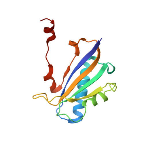
1DBF

CHORISMATE MUTASE FROM BACILLUS SUBTILIS AT 1.30 ANGSTROM


Experimental Data Snapshot

  • Method: X-RAY DIFFRACTION
  • Resolution: 1.30 Å
  • R-Value Free: 0.235 
  • R-Value Observed: 0.167 

Starting Model: experimental
View more details

wwPDB Validation   3D Report Full Report


This is version 1.4 of the entry. See complete history


Literature

The 1.30 A resolution structure of the Bacillus subtilis chorismate mutase catalytic homotrimer.

Ladner, J.E.Reddy, P.Davis, A.Tordova, M.Howard, A.J.Gilliland, G.L.

(2000) Acta Crystallogr D Biol Crystallogr 56: 673-683

  • DOI: https://doi.org/10.1107/s0907444900004625
  • Primary Citation of Related Structures:  
    1DBF

  • PubMed Abstract: 

    The crystal structure of the Bacillus subtilis chorismate mutase, an enzyme of the aromatic amino acids biosynthetic pathway, was determined to 1.30 A resolution. The structure of the homotrimer was determined by molecular replacement using orthorhombic crystals of space group P2(1)2(1)2(1) with unit-cell parameters a = 52.2, b = 83. 8, c = 86.0 A. The ABC trimer of the monoclinic crystal structure [Chook et al. (1994), J. Mol. Biol. 240, 476-500] was used as the starting model. The final coordinates are composed of three complete polypeptide chains of 127 amino-acid residues. In addition, there are nine sulfate ions, five glycerol molecules and 424 water molecules clearly visible in the structure. This structure was refined with aniosotropic temperature factors, has excellent geometry and a crystallographic R factor of 0.169 with an R(free) of 0.236. The three active sites of the macromolecule are at the subunit interfaces, with residues from two subunits contributing to each site. This orthorhombic crystal form was grown using ammonium sulfate as the precipitant; glycerol was used as a cryoprotectant during data collection. A glycerol molecule and sulfate ion in each of the active sites was found mimicking a transition-state analog. In this structure, the C-terminal tails of the subunits of the trimer are hydrogen bonded to residues of the active site of neighboring trimers in the crystal and thus cross-link the molecules in the crystal lattice.


  • Organizational Affiliation

    National Institute of Standards and Technology and the University of Maryland Biotechnology Institute, Rockville, MD 20850, USA.


Macromolecules
Find similar proteins by:  (by identity cutoff)  |  3D Structure
Entity ID: 1
MoleculeChains Sequence LengthOrganismDetailsImage
PROTEIN (CHORISMATE MUTASE)
A, B, C
127Bacillus subtilisMutation(s): 0 
EC: 5.4.99.5
UniProt
Find proteins for P19080 (Bacillus subtilis (strain 168))
Explore P19080 
Go to UniProtKB:  P19080
Entity Groups  
Sequence Clusters30% Identity50% Identity70% Identity90% Identity95% Identity100% Identity
UniProt GroupP19080
Sequence Annotations
Expand
  • Reference Sequence
Small Molecules
Ligands 2 Unique
IDChains Name / Formula / InChI Key2D Diagram3D Interactions
SO4
Query on SO4

Download Ideal Coordinates CCD File 
D [auth A]
E [auth A]
F [auth A]
G [auth A]
K [auth B]
D [auth A],
E [auth A],
F [auth A],
G [auth A],
K [auth B],
L [auth B],
M [auth B],
O [auth C],
P [auth C]
SULFATE ION
O4 S
QAOWNCQODCNURD-UHFFFAOYSA-L
GOL
Query on GOL

Download Ideal Coordinates CCD File 
H [auth A],
I [auth A],
J [auth A],
N [auth B],
Q [auth C]
GLYCEROL
C3 H8 O3
PEDCQBHIVMGVHV-UHFFFAOYSA-N
Experimental Data & Validation

Experimental Data

  • Method: X-RAY DIFFRACTION
  • Resolution: 1.30 Å
  • R-Value Free: 0.235 
  • R-Value Observed: 0.167 
  • Space Group: P 21 21 21
Unit Cell:
Length ( Å )Angle ( ˚ )
a = 52.2α = 90
b = 83.77β = 90
c = 85.96γ = 90
Software Package:
Software NamePurpose
AMoREphasing
SHELXL-97refinement
X-GENdata reduction
X-GENdata scaling

Structure Validation

View Full Validation Report


Entry History 

Deposition Data

Revision History  (Full details and data files)

  • Version 1.0: 2000-06-07
    Type: Initial release
  • Version 1.1: 2008-04-27
    Changes: Version format compliance
  • Version 1.2: 2011-07-13
    Changes: Non-polymer description, Version format compliance
  • Version 1.3: 2017-10-04
    Changes: Refinement description
  • Version 1.4: 2023-08-09
    Changes: Data collection, Database references, Derived calculations, Refinement description