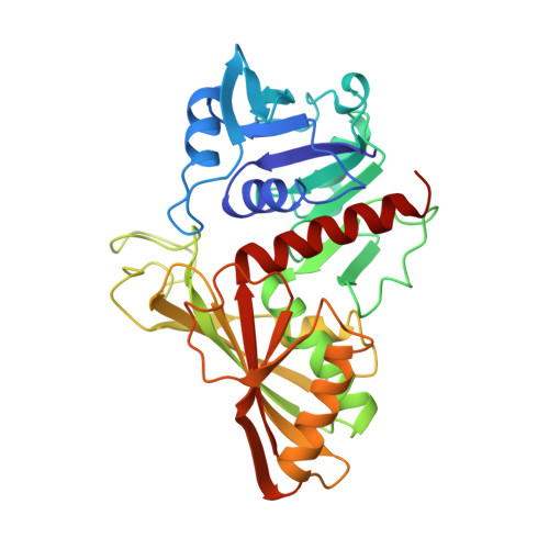
1GD1

STRUCTURE OF HOLO-GLYCERALDEHYDE-3-PHOSPHATE DEHYDROGENASE FROM BACILLUS STEAROTHERMOPHILUS AT 1.8 ANGSTROMS RESOLUTION


Experimental Data Snapshot

  • Method: X-RAY DIFFRACTION
  • Resolution: 1.80 Å
  • R-Value Observed: 0.177 

wwPDB Validation   3D Report Full Report


Ligand Structure Quality Assessment 


This is version 2.1 of the entry. See complete history


Literature

Structure of holo-glyceraldehyde-3-phosphate dehydrogenase from Bacillus stearothermophilus at 1.8 A resolution.

Skarzynski, T.Moody, P.C.Wonacott, A.J.

(1987) J Mol Biol 193: 171-187

  • DOI: https://doi.org/10.1016/0022-2836(87)90635-8
  • Primary Citation of Related Structures:  
    1GD1

  • PubMed Abstract: 

    The structure of holo-glyceraldehyde-3-phosphate dehydrogenase from Bacillus stearothermophilus has been crystallographically refined at 1.8 A resolution using restrained least-squares refinement methods. The final crystallographic R-factor for 93,120 reflexions with F greater than 3 sigma (F) is 0.177. The asymmetric unit of the crystal contains a complete tetramer, the final model of which incorporates a total of 10,272 unique protein and coenzyme atoms together with 677 bound solvent molecules. The structure has been analysed with respect to molecular symmetry, intersubunit contacts, coenzyme binding and active site geometry. The refined model shows the four independent subunits to be remarkable similar apart from local deviations due to intermolecular contacts within the crystal lattice. A number of features are revealed that had previously been misinterpreted from an earlier 2.7 A electron density map. Arginine at position 195 (previously thought to be a glycine) contributes to the formation of the anion binding sites in the active site pocket, which are involved in binding of the substrate and inorganic phosphates during catalysis. This residue seems to be structurally equivalent to the conserved Arg194 in the enzyme from other sources. In the crystal both of the anion binding sites are occupied by sulphate ions. The ND atom of the catalytically important His176 is hydrogen-bonded to the main-chain carbonyl oxygen of Ser177, thus fixing the plane of the histidine imidazole ring and preventing rotation. The analysis has revealed the presence of several internal salt-bridges stabilizing the tertiary and quaternary structure. A significant number of buried water molecules have been found that play an important role in the structural integrity of the molecule.


Macromolecules
Find similar proteins by:  (by identity cutoff)  |  3D Structure
Entity ID: 1
MoleculeChains Sequence LengthOrganismDetailsImage
HOLO-D-GLYCERALDEHYDE-3-PHOSPHATE DEHYDROGENASEA [auth O],
B [auth P],
C [auth Q],
D [auth R]
334Geobacillus stearothermophilusMutation(s): 0 
Gene Names: BACILLUS STEAROTHERMOPHILUS
EC: 1.2.1.12
UniProt
Find proteins for P00362 (Geobacillus stearothermophilus)
Explore P00362 
Go to UniProtKB:  P00362
Entity Groups  
Sequence Clusters30% Identity50% Identity70% Identity90% Identity95% Identity100% Identity
UniProt GroupP00362
Sequence Annotations
Expand
  • Reference Sequence
Small Molecules
Ligands 2 Unique
IDChains Name / Formula / InChI Key2D Diagram3D Interactions
NAD
Query on NAD

Download Ideal Coordinates CCD File 
G [auth O],
J [auth P],
M [auth Q],
P [auth R]
NICOTINAMIDE-ADENINE-DINUCLEOTIDE
C21 H27 N7 O14 P2
BAWFJGJZGIEFAR-NNYOXOHSSA-N
SO4
Query on SO4

Download Ideal Coordinates CCD File 
E [auth O]
F [auth O]
H [auth P]
I [auth P]
K [auth Q]
E [auth O],
F [auth O],
H [auth P],
I [auth P],
K [auth Q],
L [auth Q],
N [auth R],
O [auth R]
SULFATE ION
O4 S
QAOWNCQODCNURD-UHFFFAOYSA-L
Experimental Data & Validation

Experimental Data

  • Method: X-RAY DIFFRACTION
  • Resolution: 1.80 Å
  • R-Value Observed: 0.177 
  • Space Group: P 1 21 1
Unit Cell:
Length ( Å )Angle ( ˚ )
a = 82.44α = 90
b = 124.1β = 108.98
c = 82.54γ = 90
Software Package:
Software NamePurpose
PROLSQrefinement

Structure Validation

View Full Validation Report


Ligand Structure Quality Assessment 


Entry History 

Deposition Data

Revision History  (Full details and data files)

  • Version 1.0: 1988-01-16
    Type: Initial release
  • Version 1.1: 2008-03-03
    Changes: Version format compliance
  • Version 1.2: 2011-07-13
    Changes: Advisory, Version format compliance
  • Version 2.0: 2023-07-26
    Type: Remediation
    Changes: Advisory, Atomic model, Data collection, Database references, Derived calculations, Other, Refinement description
  • Version 2.1: 2023-09-06
    Changes: Data collection, Refinement description