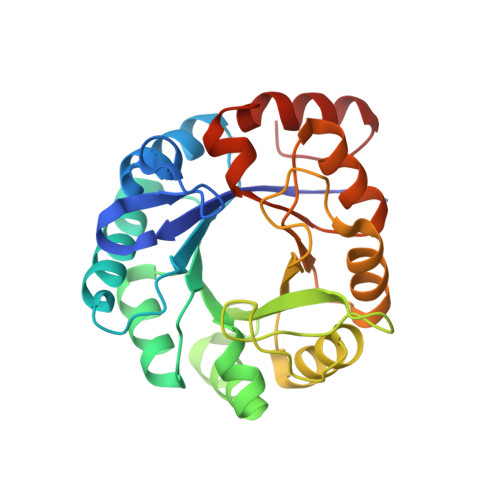
1H5Y

HisF protein from Pyrobaculum aerophilum


Experimental Data Snapshot

  • Method: X-RAY DIFFRACTION
  • Resolution: 2.00 Å
  • R-Value Free: 0.227 
  • R-Value Work: 0.206 
  • R-Value Observed: 0.207 

Starting Model: experimental
View more details

wwPDB Validation   3D Report Full Report


This is version 1.3 of the entry. See complete history


Literature

Structure of Hisf, a Histidine Biosynthetic Protein from Pyrobaculum Aerophilum

Banfield, M.J.Lott, J.S.Arcus, V.Mccarthy, A.A.Baker, E.N.

(2001) Acta Crystallogr D Biol Crystallogr 57: 1518

  • DOI: https://doi.org/10.1107/s0907444901012604
  • Primary Citation of Related Structures:  
    1H5Y

  • PubMed Abstract: 

    HisF (imidazole glycerol phosphate synthase) is an important branch-point enzyme in the histidine biosynthetic pathway of microorganisms. Because of its potential relevance for structure-based drug design, the crystal structure of HisF from the hyperthermophilic archaeon Pyrobaculum aerophilum has been determined. The structure was determined by molecular replacement and refined at 2.0 A resolution to a crystallographic R factor of 20.6% and a free R of 22.7%. The structure adopts a classic (beta/alpha)(8) barrel fold and has networks of surface salt bridges that may contribute to thermostability. The active site is marked out by the presence of two bound phosphate ions and two glycerol molecules that delineate a long groove at one end of the (beta/alpha)(8) barrel. The two phosphate ions, 17 A apart, are bound to sequence-conserved structural motifs that seem likely to provide much of the specificity for the two phosphate groups of the HisF substrate. The two glycerol molecules bind in the vicinity of other sequence-conserved residues that are likely to be involved in binding and/or catalysis. Comparisons with the homologous HisF from Thermatoga maritima reveal a displaced loop that may serve as a lid over the active site.


  • Organizational Affiliation

    School of Biological Sciences, University of Auckland, Private Bag 92019, Auckland, New Zealand.


Macromolecules
Find similar proteins by:  (by identity cutoff)  |  3D Structure
Entity ID: 1
MoleculeChains Sequence LengthOrganismDetailsImage
HISF
A, B
253Pyrobaculum aerophilumMutation(s): 0 
EC: 4.3.2.10
UniProt
Find proteins for Q8ZY16 (Pyrobaculum aerophilum (strain ATCC 51768 / DSM 7523 / JCM 9630 / CIP 104966 / NBRC 100827 / IM2))
Explore Q8ZY16 
Go to UniProtKB:  Q8ZY16
Entity Groups  
Sequence Clusters30% Identity50% Identity70% Identity90% Identity95% Identity100% Identity
UniProt GroupQ8ZY16
Sequence Annotations
Expand
  • Reference Sequence
Small Molecules
Ligands 2 Unique
IDChains Name / Formula / InChI Key2D Diagram3D Interactions
PO4
Query on PO4

Download Ideal Coordinates CCD File 
C [auth A],
D [auth A],
G [auth B],
H [auth B]
PHOSPHATE ION
O4 P
NBIIXXVUZAFLBC-UHFFFAOYSA-K
GOL
Query on GOL

Download Ideal Coordinates CCD File 
E [auth A],
F [auth A],
I [auth B],
J [auth B]
GLYCEROL
C3 H8 O3
PEDCQBHIVMGVHV-UHFFFAOYSA-N
Experimental Data & Validation

Experimental Data

  • Method: X-RAY DIFFRACTION
  • Resolution: 2.00 Å
  • R-Value Free: 0.227 
  • R-Value Work: 0.206 
  • R-Value Observed: 0.207 
  • Space Group: P 61
Unit Cell:
Length ( Å )Angle ( ˚ )
a = 105.896α = 90
b = 105.896β = 90
c = 141.828γ = 120
Software Package:
Software NamePurpose
REFMACrefinement
DENZOdata reduction
SCALEPACKdata scaling
AMoREphasing

Structure Validation

View Full Validation Report


Entry History 

Deposition Data

Revision History  (Full details and data files)

  • Version 1.0: 2001-06-01
    Type: Initial release
  • Version 1.1: 2011-05-08
    Changes: Version format compliance
  • Version 1.2: 2011-07-13
    Changes: Version format compliance
  • Version 1.3: 2023-12-13
    Changes: Data collection, Database references, Derived calculations, Other, Refinement description