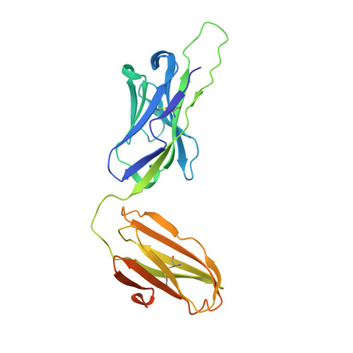
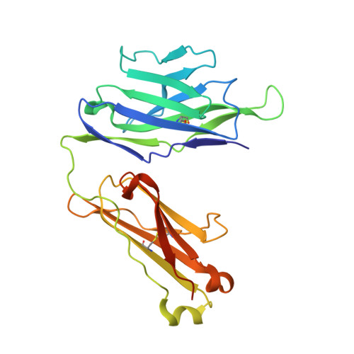
1HXM

Crystal Structure of a Human Vgamma9/Vdelta2 T Cell Receptor


Experimental Data Snapshot

  • Method: X-RAY DIFFRACTION
  • Resolution: 3.12 Å
  • R-Value Free: 0.266 
  • R-Value Work: 0.219 
  • R-Value Observed: 0.219 

wwPDB Validation   3D Report Full Report


This is version 1.3 of the entry. See complete history


Literature

Structure of a human gammadelta T-cell antigen receptor.

Allison, T.J.Winter, C.C.Fournie, J.J.Bonneville, M.Garboczi, D.N.

(2001) Nature 411: 820-824

  • DOI: https://doi.org/10.1038/35081115
  • Primary Citation of Related Structures:  
    1HXM

  • PubMed Abstract: 

    T-cell antigen receptors composed of gamma and delta polypeptide chains (gammadelta TCRs) can directly recognize antigens in the form of intact proteins or non-peptide compounds, unlike alphabeta TCRs, which recognize antigens bound to major histocompatibility complex molecules (MHC). About 5% of peripheral blood T cells bear gammadelta TCRs, most of which recognize non-peptide phosphorylated antigens. Here we describe the 3.1 A resolution structure of a human gammadelta TCR from a T-cell clone that is phosphoantigen-reactive. The orientation of the variable (V) and constant (C) regions of the gammadelta TCR is unique when compared with alphabeta TCRs or antibodies, and results from an unusually small angle between the Vgamma and Cgamma domains. The complementarity-determining regions (CDRs) of the V domains exhibit a chemically reasonable binding site for phosphorylated antigens, providing a possible explanation for the canonical usage of the Vgamma9 and Vdelta2 gene segments by phosphoantigen-reactive receptors. Although the gammadelta TCR V domains are similar in overall structure to those of alphabeta TCRs, gammadelta TCR C domains are markedly different. Structural differences in Cgamma and Cdelta, and in the location of the disulphide bond between them, may enable gammadelta TCRs to form different recognition/signalling complexes than alphabeta TCRs.


  • Organizational Affiliation

    Laboratory of Immunogenetics, National Institute of Allergy and Infectious Diseases, Rockville, Maryland 20852, USA. tallison@niaid.nih.gov


Macromolecules
Find similar proteins by:  (by identity cutoff)  |  3D Structure
Entity ID: 1
MoleculeChains Sequence LengthOrganismDetailsImage
GAMMA-DELTA T-CELL RECEPTOR
A, C, E, G
229Homo sapiensMutation(s): 0 
Gene Names: VD2DD3JD1CD
UniProt & NIH Common Fund Data Resources
Find proteins for B7Z8K6 (Homo sapiens)
Explore B7Z8K6 
Go to UniProtKB:  B7Z8K6
PHAROS:  B7Z8K6
Entity Groups  
Sequence Clusters30% Identity50% Identity70% Identity90% Identity95% Identity100% Identity
UniProt GroupB7Z8K6
Sequence Annotations
Expand
  • Reference Sequence
Find similar proteins by:  (by identity cutoff)  |  3D Structure
Entity ID: 2
MoleculeChains Sequence LengthOrganismDetailsImage
GAMMA-DELTA T-CELL RECEPTOR
B, D, F, H
242Homo sapiensMutation(s): 0 
Gene Names: VG9JGPCG1
UniProt & NIH Common Fund Data Resources
Find proteins for P03986 (Homo sapiens)
Explore P03986 
Go to UniProtKB:  P03986
PHAROS:  P03986
Entity Groups  
Sequence Clusters30% Identity50% Identity70% Identity90% Identity95% Identity100% Identity
UniProt GroupP03986
Sequence Annotations
Expand
  • Reference Sequence
Experimental Data & Validation

Experimental Data

  • Method: X-RAY DIFFRACTION
  • Resolution: 3.12 Å
  • R-Value Free: 0.266 
  • R-Value Work: 0.219 
  • R-Value Observed: 0.219 
  • Space Group: P 1 21 1
Unit Cell:
Length ( Å )Angle ( ˚ )
a = 74.048α = 90
b = 151.736β = 99.48
c = 97.873γ = 90
Software Package:
Software NamePurpose
SCALEPACKdata scaling
SHARPphasing
CNSrefinement

Structure Validation

View Full Validation Report


Entry History 

Deposition Data

Revision History  (Full details and data files)

  • Version 1.0: 2001-06-20
    Type: Initial release
  • Version 1.1: 2008-04-27
    Changes: Version format compliance
  • Version 1.2: 2011-07-13
    Changes: Version format compliance
  • Version 1.3: 2017-10-04
    Changes: Refinement description