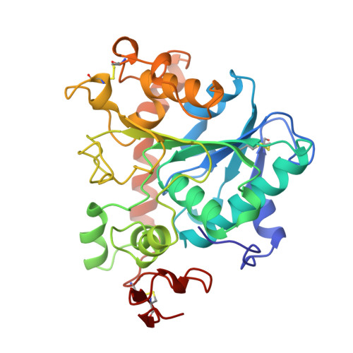
1LBT

LIPASE (E.C.3.1.1.3) (TRIACYLGLYCEROL HYDROLASE)


Experimental Data Snapshot

  • Method: X-RAY DIFFRACTION
  • Resolution: 2.50 Å
  • R-Value Free: 0.193 
  • R-Value Work: 0.172 
  • R-Value Observed: 0.172 

wwPDB Validation   3D Report Full Report


Ligand Structure Quality Assessment 


This is version 2.1 of the entry. See complete history


Literature

Crystallographic and molecular-modeling studies of lipase B from Candida antarctica reveal a stereospecificity pocket for secondary alcohols.

Uppenberg, J.Ohrner, N.Norin, M.Hult, K.Kleywegt, G.J.Patkar, S.Waagen, V.Anthonsen, T.Jones, T.A.

(1995) Biochemistry 34: 16838-16851

  • DOI: https://doi.org/10.1021/bi00051a035
  • Primary Citation of Related Structures:  
    1LBS, 1LBT

  • PubMed Abstract: 

    Many lipases are potent catalysts of stereoselective reactions and are therefore of interest for use in chemical synthesis. The crystal structures of lipases show a large variation in the shapes of their active site environments that may explain the large variation in substrate specificity of these enzymes. We have determined the three-dimensional structure of Candida antarctica lipase B (CALB) cocrystallized with the detergent Tween 80. In another crystal form, the structure of the enzyme in complex with a covalently bound phosphonate inhibitor has been determined. In both structures, the active site is exposed to the external solvent. The potential lid-forming helix alpha 5 in CALB is well-ordered in the Tween 80 structure and disordered in the inhibitor complex. The tetrahedral intermediates of two chiral substrates have been modeled on the basis of available structural and biochemical information. The results of this study provide a structural explanation for the high stereoselectivity of CALB toward many secondary alcohols.


  • Organizational Affiliation

    Department of Molecular Biology, Uppsala University, Sweden.


Macromolecules
Find similar proteins by:  (by identity cutoff)  |  3D Structure
Entity ID: 1
MoleculeChains Sequence LengthOrganismDetailsImage
LIPASE B
A, B
317Moesziomyces antarcticusMutation(s): 0 
EC: 3.1.1.3
UniProt
Find proteins for P41365 (Pseudozyma antarctica)
Explore P41365 
Go to UniProtKB:  P41365
Entity Groups  
Sequence Clusters30% Identity50% Identity70% Identity90% Identity95% Identity100% Identity
UniProt GroupP41365
Glycosylation
Glycosylation Sites: 1
Sequence Annotations
Expand
  • Reference Sequence
Oligosaccharides

Help

Entity ID: 2
MoleculeChains Length2D Diagram Glycosylation3D Interactions
2-acetamido-2-deoxy-beta-D-glucopyranose-(1-4)-2-acetamido-2-deoxy-beta-D-glucopyranose
C, D
2N-Glycosylation
Glycosylation Resources
GlyTouCan:  G42666HT
GlyCosmos:  G42666HT
GlyGen:  G42666HT
Experimental Data & Validation

Experimental Data

  • Method: X-RAY DIFFRACTION
  • Resolution: 2.50 Å
  • R-Value Free: 0.193 
  • R-Value Work: 0.172 
  • R-Value Observed: 0.172 
  • Space Group: P 1 21 1
Unit Cell:
Length ( Å )Angle ( ˚ )
a = 95.1α = 90
b = 50.2β = 90.6
c = 99.5γ = 90
Software Package:
Software NamePurpose
MARKdata collection
SDMSdata collection
X-PLORmodel building
X-PLORrefinement
MARKdata reduction
SDMSdata reduction
X-PLORphasing

Structure Validation

View Full Validation Report


Ligand Structure Quality Assessment 


Entry History 

Deposition Data

Revision History  (Full details and data files)

  • Version 1.0: 1995-10-15
    Type: Initial release
  • Version 1.1: 2008-03-03
    Changes: Version format compliance
  • Version 1.2: 2011-07-13
    Changes: Non-polymer description, Version format compliance
  • Version 1.3: 2017-11-29
    Changes: Derived calculations, Other
  • Version 2.0: 2020-07-29
    Type: Remediation
    Reason: Carbohydrate remediation
    Changes: Atomic model, Data collection, Derived calculations, Structure summary
  • Version 2.1: 2024-10-30
    Changes: Data collection, Database references, Structure summary