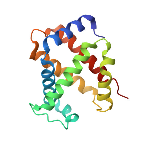
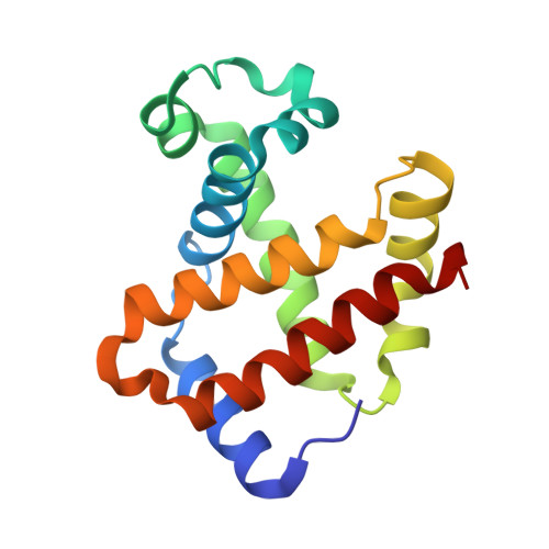
1LJW

Crystal Structure of Human Carbonmonoxy Hemoglobin at 2.16 A: A Snapshot of the Allosteric Transition


Experimental Data Snapshot

  • Method: X-RAY DIFFRACTION
  • Resolution: 2.16 Å
  • R-Value Free: 0.259 
  • R-Value Work: 0.194 
  • R-Value Observed: 0.205 

Starting Model: experimental
View more details

wwPDB Validation   3D Report Full Report


Ligand Structure Quality Assessment 


This is version 1.4 of the entry. See complete history


Literature

Structure of human carbonmonoxyhemoglobin at 2.16 A: a snapshot of the allosteric transition.

Safo, M.K.Burnett, J.C.Musayev, F.N.Nokuri, S.Abraham, D.J.

(2002) Acta Crystallogr D Biol Crystallogr 58: 2031-2037

  • DOI: https://doi.org/10.1107/s0907444902015809
  • Primary Citation of Related Structures:  
    1LJW

  • PubMed Abstract: 

    A 2.16 A resolution structure of high-salt human carbonmonoxyhemoglobin crystallized at pH 6.4 is reported. The quaternary structure is similar to that of 'classic' R-state hemoglobin; however, subtle but significant tertiary structural changes are observed at the alpha(1)beta(2) and symmetrically equivalent alpha(2)beta(1) interfaces--these are the key subunit interfaces that govern the allosteric transition between the R and T states. Specifically, the movement and weakening of two important hydrogen bonds that are diagnostic for R-state structures, beta(2)His97-alpha(1)Thr38 and beta(2)Arg40-alpha(1)Thr41, have been observed. In addition, a phosphate molecule bound between the two beta-subunits (at the entrance to the central water cavity) has been identified and electron density indicates that this molecule occupies two alternate positions that are related by the dyad axis. Both positions superimpose on the 2,3-diphosphoglycerate binding site. One phosphate conformer interacts with beta(2)Asn139, beta(1)His143 and beta(1)His146, while the second interacts with symmetry-related counterparts (beta(1)Asn139, beta(2)His143 and beta(2)His146).


  • Organizational Affiliation

    School of Pharmacy, Department of Medicinal Chemistry and Institute for Structural Biology and Drug Discovery, Virginia Commonwealth University, Richmond, VA 23219, USA. msafo@hsc.vcu.edu


Macromolecules
Find similar proteins by:  (by identity cutoff)  |  3D Structure
Entity ID: 1
MoleculeChains Sequence LengthOrganismDetailsImage
hemoglobin alpha chain141Homo sapiensMutation(s): 0 
UniProt & NIH Common Fund Data Resources
Find proteins for P69905 (Homo sapiens)
Explore P69905 
Go to UniProtKB:  P69905
PHAROS:  P69905
Entity Groups  
Sequence Clusters30% Identity50% Identity70% Identity90% Identity95% Identity100% Identity
UniProt GroupP69905
Sequence Annotations
Expand
  • Reference Sequence
Find similar proteins by:  (by identity cutoff)  |  3D Structure
Entity ID: 2
MoleculeChains Sequence LengthOrganismDetailsImage
hemoglobin beta chain146Homo sapiensMutation(s): 0 
UniProt & NIH Common Fund Data Resources
Find proteins for P68871 (Homo sapiens)
Explore P68871 
Go to UniProtKB:  P68871
PHAROS:  P68871
GTEx:  ENSG00000244734 
Entity Groups  
Sequence Clusters30% Identity50% Identity70% Identity90% Identity95% Identity100% Identity
UniProt GroupP68871
Sequence Annotations
Expand
  • Reference Sequence
Experimental Data & Validation

Experimental Data

  • Method: X-RAY DIFFRACTION
  • Resolution: 2.16 Å
  • R-Value Free: 0.259 
  • R-Value Work: 0.194 
  • R-Value Observed: 0.205 
  • Space Group: P 41 21 2
Unit Cell:
Length ( Å )Angle ( ˚ )
a = 53.566α = 90
b = 53.566β = 90
c = 192.96γ = 90
Software Package:
Software NamePurpose
bioteXdata collection
bioteXdata reduction
CNSrefinement
bioteXdata scaling
CNSphasing

Structure Validation

View Full Validation Report


Ligand Structure Quality Assessment 


Entry History 

Deposition Data

Revision History  (Full details and data files)

  • Version 1.0: 2002-05-01
    Type: Initial release
  • Version 1.1: 2008-04-28
    Changes: Version format compliance
  • Version 1.2: 2011-07-13
    Changes: Derived calculations, Version format compliance
  • Version 1.3: 2017-10-11
    Changes: Refinement description
  • Version 1.4: 2023-08-16
    Changes: Data collection, Database references, Derived calculations, Refinement description