1M9Z | pdb_00001m9z

CRYSTAL STRUCTURE OF HUMAN TGF-BETA TYPE II RECEPTOR LIGAND BINDING DOMAIN


Experimental Data Snapshot

  • Method: X-RAY DIFFRACTION
  • Resolution: 1.05 Å
  • R-Value Free: 
    0.166 (Depositor) 
  • R-Value Work: 
    0.156 (Depositor) 
  • R-Value Observed: 
    0.156 (Depositor) 

wwPDB Validation 3D Report Full Report

Validation slider image for 1M9Z

This is version 1.5 of the entry. See complete history

Literature

THE 1.1A CRYSTAL STRUCTURE OF HUMAN TGF-BETA TYPE II RECEPTOR LIGAND BINDING DOMAIN

Boesen, C.C.Radaev, S.Motyka, S.A.Patamawenu, A.Sun, P.D.

(2002) Structure 10: 913-919

  • DOI: https://doi.org/10.1016/s0969-2126(02)00780-3
  • Primary Citation Related Structures: 
    1M9Z

  • PubMed Abstract: 

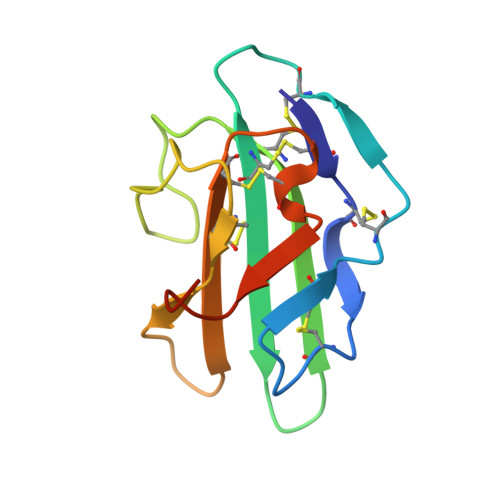
    Transforming growth factor beta (TGF-beta) is involved in a wide range of biological functions including development, carcinogenesis, and immune regulation. Here we report the 1.1 A resolution crystal structure of human TGF-beta type II receptor ectodomain (TBRII). The overall structure of TBRII is similar to that of activin type II receptor ectodomain (ActRII) and bone morphogenic protein receptor type IA (BRIA). It displays a three-finger toxin fold with fingers formed by the beta strand pairs beta1-beta2, beta3-beta4, and beta5-beta6. The first finger in the TBRII is significantly longer than in ActRII and BRIA and folds tightly between the second finger and the C terminus. Surface charge distributions and hydrophobic patches predict potential TBRII binding sites.


  • Organizational Affiliation
    • Structural Biology Section, Laboratory of Immunogenetics, National Institute of Allergy and Infectious Diseases, National Institutes of Health, Rockville, MD 20852, USA.

Macromolecule Content 

  • Total Structure Weight: 12.72 kDa 
  • Atom Count: 1,011 
  • Modeled Residue Count: 105 
  • Deposited Residue Count: 111 
  • Unique protein chains: 1

Macromolecules

Find similar proteins by:|  3D Structure
Entity ID: 1
MoleculeChains  Sequence LengthOrganismDetailsImage
TGF-BETA RECEPTOR TYPE II111Homo sapiensMutation(s): 2 
EC: 2.7.11.30
UniProt & NIH Common Fund Data Resources
Find proteins for P37173 (Homo sapiens)
Explore P37173 
Go to UniProtKB:  P37173
PHAROS:  P37173
GTEx:  ENSG00000163513 
Entity Groups
Sequence Clusters30% Identity50% Identity70% Identity90% Identity95% Identity100% Identity
UniProt GroupP37173
Sequence Annotations
Expand
Reference Sequence

Small Molecules

Ligands 1 Unique
IDChains Name / Formula / InChI Key2D Diagram3D Interactions
GOL

Query on GOL


Download:Ideal Coordinates CCD File
B [auth A]GLYCEROL
C3 H8 O3
PEDCQBHIVMGVHV-UHFFFAOYSA-N

Experimental Data & Validation

Experimental Data

  • Method: X-RAY DIFFRACTION
  • Resolution: 1.05 Å
  • R-Value Free:  0.166 (Depositor) 
  • R-Value Work:  0.156 (Depositor) 
  • R-Value Observed: 0.156 (Depositor) 
Space Group: P 21 21 21
Unit Cell:
Length ( Å )Angle ( ˚ )
a = 35.471α = 90
b = 40.658β = 90
c = 76.154γ = 90
Software Package:
Software NamePurpose
REFMACrefinement
SCALEPACKdata scaling
CNSrefinement
HKL-2000data collection
HKL-2000data reduction
CNSphasing

Structure Validation

View Full Validation Report


Entry History 

Deposition Data

Revision History  (Full details and data files)

  • Version 1.0: 2002-09-11
    Type: Initial release
  • Version 1.1: 2008-04-28
    Changes: Version format compliance
  • Version 1.2: 2011-07-13
    Changes: Non-polymer description, Version format compliance
  • Version 1.3: 2019-07-24
    Changes: Advisory, Data collection, Refinement description
  • Version 1.4: 2021-10-27
    Changes: Advisory, Database references, Derived calculations
  • Version 1.5: 2024-10-16
    Changes: Data collection, Structure summary