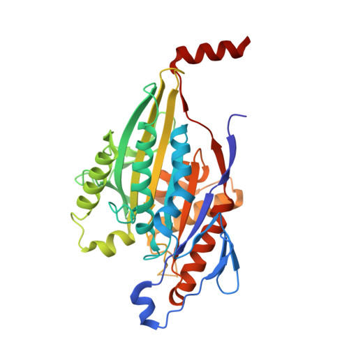
1MKJ

Human Kinesin Motor Domain With Docked Neck Linker


Experimental Data Snapshot

  • Method: X-RAY DIFFRACTION
  • Resolution: 2.70 Å
  • R-Value Free: 0.255 
  • R-Value Work: 0.209 
  • R-Value Observed: 0.209 

Starting Model: experimental
View more details

wwPDB Validation   3D Report Full Report


Ligand Structure Quality Assessment 


This is version 1.3 of the entry. See complete history


Literature

Two conformations in the human kinesin power stroke defined by X-ray crystallography and EPR spectroscopy.

Sindelar, C.V.Budny, M.J.Rice, S.Naber, N.Fletterick, R.Cooke, R.

(2002) Nat Struct Biol 9: 844-848

  • DOI: https://doi.org/10.1038/nsb852
  • Primary Citation of Related Structures:  
    1MKJ

  • PubMed Abstract: 

    Crystal structures of the molecular motor kinesin show conformational variability in a structural element called the neck linker. Conformational change in the neck linker, initiated by ATP exchange, is thought to drive the movement of kinesin along the microtubule track. We use site-specific EPR measurements to show that when microtubules are absent, the neck linker exists in equilibrium between two structural states (disordered and 'docked'). The active site nucleotide does not control the position taken by the neck linker. However, we find that sulfate can specifically bind near the nucleotide site and stabilize the docked neck linker conformation, which we confirmed by solving a new crystal structure. Comparing the crystal structures of our construct with the docked or undocked neck linker reveals how microtubule binding may activate the nucleotide-sensing mechanism of kinesin, allowing neck linker transitions to power motility.


  • Organizational Affiliation

    Department of Biochemistry and Biophysics, University of California, San Francisco, California 94143, USA.


Macromolecules
Find similar proteins by:  (by identity cutoff)  |  3D Structure
Entity ID: 1
MoleculeChains Sequence LengthOrganismDetailsImage
KINESIN HEAVY CHAIN349Homo sapiensMutation(s): 0 
UniProt & NIH Common Fund Data Resources
Find proteins for P33176 (Homo sapiens)
Explore P33176 
Go to UniProtKB:  P33176
PHAROS:  P33176
GTEx:  ENSG00000170759 
Entity Groups  
Sequence Clusters30% Identity50% Identity70% Identity90% Identity95% Identity100% Identity
UniProt GroupP33176
Sequence Annotations
Expand
  • Reference Sequence
Experimental Data & Validation

Experimental Data

  • Method: X-RAY DIFFRACTION
  • Resolution: 2.70 Å
  • R-Value Free: 0.255 
  • R-Value Work: 0.209 
  • R-Value Observed: 0.209 
  • Space Group: P 21 21 21
Unit Cell:
Length ( Å )Angle ( ˚ )
a = 73.796α = 90
b = 74.086β = 90
c = 91.543γ = 90
Software Package:
Software NamePurpose
HKL-2000data collection
SCALEPACKdata scaling
CNSrefinement
HKL-2000data reduction
CNSphasing

Structure Validation

View Full Validation Report


Ligand Structure Quality Assessment 


Entry History 

Deposition Data

Revision History  (Full details and data files)

  • Version 1.0: 2002-10-30
    Type: Initial release
  • Version 1.1: 2008-04-28
    Changes: Version format compliance
  • Version 1.2: 2011-07-13
    Changes: Version format compliance
  • Version 1.3: 2024-02-14
    Changes: Data collection, Database references, Derived calculations, Refinement description