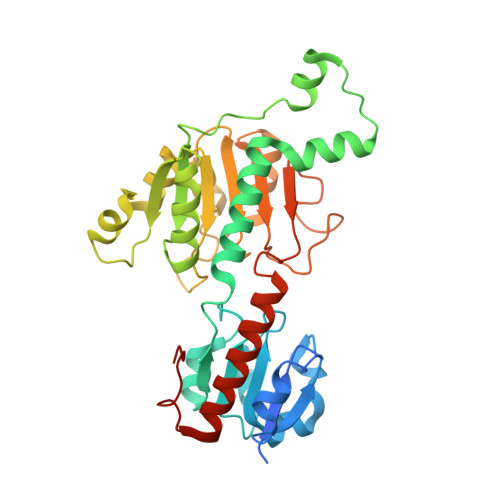
1MX3

Crystal structure of CtBP dehydrogenase core holo form


Experimental Data Snapshot

  • Method: X-RAY DIFFRACTION
  • Resolution: 1.95 Å
  • R-Value Free: 0.253 
  • R-Value Work: 0.211 

wwPDB Validation   3D Report Full Report


Ligand Structure Quality Assessment 


This is version 1.3 of the entry. See complete history


Literature

Transcription Corepressor CtBP Is an NAD+-Regulated Dehydrogenase

Kumar, V.Carlson, J.E.Ohgi, K.E.Edwards, T.E.Rose, D.W.Escalante, C.R.Aggarwal, A.K.

(2002) Mol Cell 10: 857-869

  • DOI: https://doi.org/10.1016/s1097-2765(02)00650-0
  • Primary Citation of Related Structures:  
    1MX3

  • PubMed Abstract: 

    Transcriptional repression is based on the selective actions of recruited corepressor complexes, including those with enzymatic activities. One well-characterized developmentally important corepressor is the C-terminal binding protein (CtBP). Although intriguingly related in sequence to D2 hydroxyacid dehydrogenases, the mechanism by which CtBP functions remains unclear. We report here biochemical and crystallographic studies which reveal that CtBP is a functional dehydrogenase. In addition, both a cofactor-dependent conformational change, with NAD(+) and NADH being equivalently effective, and the active site residues are linked to the binding of the PXDLS consensus recognition motif on repressors, such as E1A and RIP140. Together, our data suggest that CtBP is an NAD(+)-regulated component of critical complexes for specific repression events in cells.


  • Organizational Affiliation

    Department of Biology Graduate Program, University of California, San Diego, La Jolla, CA 92093, USA.


Macromolecules
Find similar proteins by:  (by identity cutoff)  |  3D Structure
Entity ID: 1
MoleculeChains Sequence LengthOrganismDetailsImage
C-terminal binding protein 1347Homo sapiensMutation(s): 0 
Gene Names: CTBP1
EC: 1.1.1
UniProt & NIH Common Fund Data Resources
Find proteins for Q13363 (Homo sapiens)
Explore Q13363 
Go to UniProtKB:  Q13363
PHAROS:  Q13363
GTEx:  ENSG00000159692 
Entity Groups  
Sequence Clusters30% Identity50% Identity70% Identity90% Identity95% Identity100% Identity
UniProt GroupQ13363
Sequence Annotations
Expand
  • Reference Sequence
Small Molecules
Ligands 2 Unique
IDChains Name / Formula / InChI Key2D Diagram3D Interactions
NAD
Query on NAD

Download Ideal Coordinates CCD File 
B [auth A]NICOTINAMIDE-ADENINE-DINUCLEOTIDE
C21 H27 N7 O14 P2
BAWFJGJZGIEFAR-NNYOXOHSSA-N
ACY
Query on ACY

Download Ideal Coordinates CCD File 
C [auth A]ACETIC ACID
C2 H4 O2
QTBSBXVTEAMEQO-UHFFFAOYSA-N
Experimental Data & Validation

Experimental Data

  • Method: X-RAY DIFFRACTION
  • Resolution: 1.95 Å
  • R-Value Free: 0.253 
  • R-Value Work: 0.211 
  • Space Group: P 64 2 2
Unit Cell:
Length ( Å )Angle ( ˚ )
a = 89.1α = 90
b = 89.1β = 90
c = 164γ = 120
Software Package:
Software NamePurpose
CNSrefinement
DENZOdata reduction
SCALEPACKdata scaling
CNSphasing

Structure Validation

View Full Validation Report


Ligand Structure Quality Assessment 


Entry History 

Deposition Data

Revision History  (Full details and data files)

  • Version 1.0: 2002-12-18
    Type: Initial release
  • Version 1.1: 2008-04-28
    Changes: Version format compliance
  • Version 1.2: 2011-07-13
    Changes: Derived calculations, Version format compliance
  • Version 1.3: 2024-02-14
    Changes: Data collection, Database references, Derived calculations