1N59

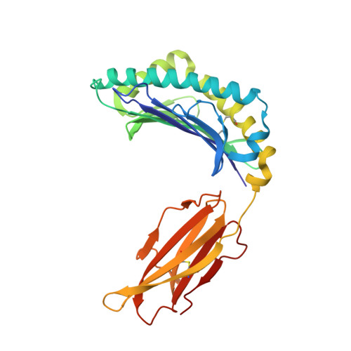
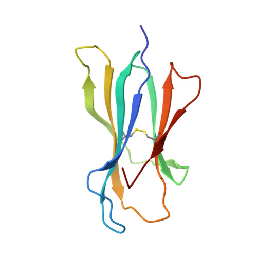
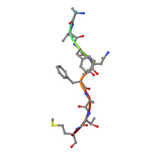
Crystal structure of the Murine class I Major Histocompatibility Complex of H-2KB, B2-Microglobulin, and A 9-Residue immunodominant peptide epitope gp33 derived from LCMV


Experimental Data Snapshot

  • Method: X-RAY DIFFRACTION
  • Resolution: 2.95 Å
  • R-Value Free: 0.287 
  • R-Value Work: 0.229 
  • R-Value Observed: 0.232 

Starting Model: experimental
View more details

wwPDB Validation   3D Report Full Report


This is version 1.5 of the entry. See complete history


Literature

A Structural Basis for LCMV Immune Evasion. Subversion of H-2D(b) and H-2K(b) Presentation of gp33 Revealed by Comparative Crystal Structure Analyses.

Achour, A.Michaelsson, J.Harris, R.A.Odeberg, J.Grufman, P.Sandberg, J.K.Levitsky, V.Karre, K.Sandalova, T.Schneider, G.

(2002) Immunity 17: 757-768

  • DOI: https://doi.org/10.1016/s1074-7613(02)00478-8
  • Primary Citation of Related Structures:  
    1N59, 1N5A

  • PubMed Abstract: 

    LCMV infection of H-2(b) mice generates a CD8(+) CTL response mainly directed toward three immunodominant epitopes. One of these, gp33, is presented by both H-2D(b) and H-2K(b) MHC class I molecules. The virus can escape immune recognition in the context of both these MHC class I molecules through single mutations of the peptide. In order to understand the underlying structural mechanism, we determined the crystal structures of both complexes. The structures reveal that the peptide is presented in two diametrically opposed manners by H-2D(b) and H-2K(b), with residues used as anchor positions in one MHC class I molecule interacting with the TCR in the other. Importantly, the peptide's N-terminal residue p1K protrudes from the binding cleft in H-2K(b). We present structural evidence that explains the functional consequences of single mutations found in escape variants.


  • Organizational Affiliation

    Microbiology and Tumor Biology Center, Karolinska Institutet, Royal School of Technology, S-106 91 Stockholm, Sweden. adnane.achour@mtc.ki.se


Macromolecules
Find similar proteins by:  (by identity cutoff)  |  3D Structure
Entity ID: 1
MoleculeChains Sequence LengthOrganismDetailsImage
H-2 class I histocompatibility antigen, K-B alpha chainA,
D [auth C]
276Mus musculusMutation(s): 0 
Gene Names: H2-K
UniProt
Find proteins for P01901 (Mus musculus)
Explore P01901 
Go to UniProtKB:  P01901
Entity Groups  
Sequence Clusters30% Identity50% Identity70% Identity90% Identity95% Identity100% Identity
UniProt GroupP01901
Sequence Annotations
Expand
  • Reference Sequence
Find similar proteins by:  (by identity cutoff)  |  3D Structure
Entity ID: 2
MoleculeChains Sequence LengthOrganismDetailsImage
Beta-2-microglobulinB,
E [auth D]
99Mus musculusMutation(s): 0 
Gene Names: B2M
UniProt & NIH Common Fund Data Resources
Find proteins for P01887 (Mus musculus)
Explore P01887 
Go to UniProtKB:  P01887
IMPC:  MGI:88127
Entity Groups  
Sequence Clusters30% Identity50% Identity70% Identity90% Identity95% Identity100% Identity
UniProt GroupP01887
Sequence Annotations
Expand
  • Reference Sequence

Find similar proteins by:  Sequence   |   3D Structure  

Entity ID: 3
MoleculeChains Sequence LengthOrganismDetailsImage
nonameric peptide, gp33 derived from lymphocytic choriomeningitis virusC [auth P],
F [auth Q]
9N/AMutation(s): 1 
UniProt
Find proteins for Q9QDK7 (Mammarenavirus choriomeningitidis)
Explore Q9QDK7 
Go to UniProtKB:  Q9QDK7
Entity Groups  
UniProt GroupQ9QDK7
Sequence Annotations
Expand
  • Reference Sequence
Experimental Data & Validation

Experimental Data

  • Method: X-RAY DIFFRACTION
  • Resolution: 2.95 Å
  • R-Value Free: 0.287 
  • R-Value Work: 0.229 
  • R-Value Observed: 0.232 
  • Space Group: P 1 21 1
Unit Cell:
Length ( Å )Angle ( ˚ )
a = 50.551α = 90
b = 88.574β = 93.72
c = 120.09γ = 90
Software Package:
Software NamePurpose
DENZOdata reduction
SCALEPACKdata scaling
AMoREphasing
REFMACrefinement

Structure Validation

View Full Validation Report


Entry History 

Deposition Data

Revision History  (Full details and data files)

  • Version 1.0: 2003-01-07
    Type: Initial release
  • Version 1.1: 2008-04-28
    Changes: Version format compliance
  • Version 1.2: 2011-07-13
    Changes: Advisory, Version format compliance
  • Version 1.3: 2018-03-07
    Changes: Data collection
  • Version 1.4: 2021-10-27
    Changes: Database references
  • Version 1.5: 2024-10-30
    Changes: Data collection, Refinement description, Structure summary