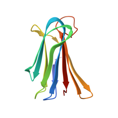
RCSB PDB - 1OUR: LecB (PA-LII) in complex with mannose

 1OUR

LecB (PA-LII) in complex with mannose


Experimental Data Snapshot

  • Method: X-RAY DIFFRACTION
  • Resolution: 1.42 Å
  • R-Value Free: 0.197 
  • R-Value Work: 0.182 
  • R-Value Observed: 0.183 

wwPDB Validation   3D Report Full Report


Ligand Structure Quality Assessment 

Created with Raphaël 2.3.0Worse 01 BetterLigand structure goodness of fit to experimental dataBest fitted MANClick on this verticalbar to view details

This is version 1.4 of the entry. See complete history


Literature

Structural Basis of Carbohydrate Recognition by the Lectin LecB from Pseudomonas aeruginosa

Loris, R.Tielker, D.Jaeger, K.-E.Wyns, L.

(2003) J Mol Biol 331: 861-870

  • DOI: https://doi.org/10.1016/s0022-2836(03)00754-x
  • Primary Citation of Related Structures:  
    1OUR, 1OUS, 1OUX, 1OVP, 1OVS, 1OXC

  • PubMed Abstract: 

    The crystal structure of Pseudomonas aeruginosa fucose-specific lectin LecB was determined in its metal-bound and metal-free state as well as in complex with fucose, mannose and fructopyranose. All three monosaccharides bind isosterically via direct interactions with two calcium ions as well as direct hydrogen bonds with several side-chains. The higher affinity for fucose is explained by the details of the binding site around C6 and O1 of fucose. In the mannose and fructose complexes, a carboxylate oxygen atom and one or two hydroxyl groups are partly shielded from solvent upon sugar binding, preventing them from completely fulfilling their hydrogen bonding potential. In the fucose complex, no such defects are observed. Instead, C6 makes favourable interactions with a small hydrophobic patch. Upon demetallization, the C terminus as well as the otherwise rigid metal-binding loop become more mobile and adopt multiple conformations.


  • Organizational Affiliation

    Vrije Universiteit Brussel, Laboratorium voor Ultrastructuur Instituut voor Moleculaire Biologie, Gebouw E, Pleinlaan 2, B-1050 Brussel, Belgium. reloris@vub.ac.be


Macromolecules
Find similar proteins by:  (by identity cutoff)  |  3D Structure
Entity ID: 1
MoleculeChains Sequence LengthOrganismDetailsImage
hypothetical protein LecB114Pseudomonas aeruginosaMutation(s): 0 
Gene Names: LecB
UniProt
Find proteins for Q9HYN5 (Pseudomonas aeruginosa (strain ATCC 15692 / DSM 22644 / CIP 104116 / JCM 14847 / LMG 12228 / 1C / PRS 101 / PAO1))
Explore Q9HYN5 
Go to UniProtKB:  Q9HYN5
Entity Groups  
Sequence Clusters30% Identity50% Identity70% Identity90% Identity95% Identity100% Identity
UniProt GroupQ9HYN5
Sequence Annotations
Expand
  • Reference Sequence
Experimental Data & Validation

Experimental Data

  • Method: X-RAY DIFFRACTION
  • Resolution: 1.42 Å
  • R-Value Free: 0.197 
  • R-Value Work: 0.182 
  • R-Value Observed: 0.183 
  • Space Group: I 2 2 2
Unit Cell:
Length ( Å )Angle ( ˚ )
a = 52.777α = 90
b = 69.184β = 90
c = 74.031γ = 90
Software Package:
Software NamePurpose
DENZOdata reduction
SCALEPACKdata scaling
AMoREphasing
CNSrefinement

Structure Validation

View Full Validation Report


Ligand Structure Quality Assessment 

Created with Raphaël 2.3.0Worse 01 BetterLigand structure goodness of fit to experimental dataBest fitted MANClick on this verticalbar to view details

Entry History 

Deposition Data

Revision History  (Full details and data files)

  • Version 1.0: 2003-09-09
    Type: Initial release
  • Version 1.1: 2008-04-29
    Changes: Version format compliance
  • Version 1.2: 2011-07-13
    Changes: Version format compliance
  • Version 1.3: 2020-07-29
    Type: Remediation
    Reason: Carbohydrate remediation
    Changes: Advisory, Data collection, Derived calculations, Structure summary
  • Version 1.4: 2024-03-13
    Changes: Data collection, Database references, Structure summary