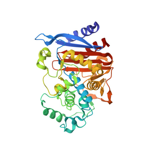
Thermodynamic cycle analysis and inhibitor design against beta-lactamase.
Roth, T.A., Minasov, G., Morandi, S., Prati, F., Shoichet, B.K.(2003) Biochemistry 42: 14483-14491
- PubMed: 14661960
- DOI: https://doi.org/10.1021/bi035054a
- Primary Citation of Related Structures:
1PI4, 1PI5 - PubMed Abstract:
Beta-lactamases are the most widespread resistance mechanism to beta-lactam antibiotics, such as the penicillins and cephalosporins. Transition-state analogues that bind to the enzymes with nanomolar affinities have been introduced in an effort to reverse the resistance conferred by these enzymes. To understand the origins of this affinity, and to guide design of future inhibitors, double-mutant thermodynamic cycle experiments were undertaken. An unexpected hydrogen bond between the nonconserved Asn289 and a key inhibitor carboxylate was observed in the X-ray crystal structure of a 1 nM inhibitor (compound 1) in complex with AmpC beta-lactamase. To investigate the energy of this hydrogen bond, the mutant enzyme N289A was made, as was an analogue of 1 that lacked the carboxylate (compound 2). The differential affinity of the four different protein and analogue complexes indicates that the carboxylate-amide hydrogen bond contributes 1.7 kcal/mol to overall binding affinity. Synthesis of an analogue of 1 where the carboxylate was replaced with an aldehyde led to an inhibitor that lost all this hydrogen bond energy, consistent with the importance of the ionic nature of this hydrogen bond. To investigate the structural bases of these energies, X-ray crystal structures of N289A/1 and N289A/2 were determined to 1.49 and 1.39 A, respectively. These structures suggest that no significant rearrangement occurs in the mutant versus the wild-type complexes with both compounds. The mutant enzymes L119A and L293A were made to investigate the interaction between a phenyl ring in 1 and these residues. Whereas deletion of the phenyl itself diminishes affinity by 5-fold, the double-mutant cycles suggest that this energy does not come through interaction with the leucines, despite the close contact in the structure. The energies of these interactions provide key information for the design of improved inhibitors against beta-lactamases. The high magnitude of the ion-dipole interaction between Asn289 and the carboxylate of 1 is consistent with the idea that ionic interactions can provide significant net affinity in inhibitor complexes.
- Department of Pharmaceutical Chemistry, University of California San Francisco, 600 16th Street, San Francisco, California 94143-2240, USA.
Organizational Affiliation:


