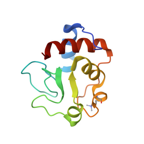
The structure and function of omega loop A replacements in cytochrome c.
Murphy, M.E., Fetrow, J.S., Burton, R.E., Brayer, G.D.(1993) Protein Sci 2: 1429-1440
- PubMed: 8401228
- DOI: https://doi.org/10.1002/pro.5560020907
- Primary Citation of Related Structures:
1RAP, 1RAQ - PubMed Abstract:
The structural and functional consequences of replacing omega-loop A (residues 18-32) in yeast iso-1-cytochrome c with the corresponding loop of Rhodospirillum rubrum cytochrome c2 have been examined. The three-dimensional structure of this loop replacement mutant RepA2 cytochrome c, and a second mutant RepA2(Val 20) cytochrome c in which residue 20 was back substituted to valine, were determined using X-ray diffraction techniques. A change in the molecular packing is evident in the RepA2 mutant protein, which has a phenylalanine at position 20, a residue considerably larger than the valine found in wild-type yeast iso-1-cytochrome c. The side chain of Phe 20 is redirected toward the molecular surface, altering the packing of this region of omega-loop A with the hydrophobic core of the protein. In the RepA2(Val 20) structure, omega-loop A contains a valine at position 20, which restores the original wild-type packing arrangement of the hydrophobic core. Also, as a result of omega-loop A replacement, residue 26 is changed from a histidine to asparagine, which results in displacements of the main-chain atoms near residue 44 to which residue 26 is hydrogen bonded. In vivo studies of the growth rate of the mutant strains on nonfermentable media indicate that the RepA2(Val 20) cytochrome c behaves much like the wild-type yeast iso-1 protein, whereas the stability and function of the RepA2 cytochrome c showed a temperature dependence. The midpoint reduction potential measured by cyclic voltammetry of the RepA2 mutant is 271 mV at 25 degrees C. This is 19 mV less than the wild-type and RepA2(Val 20) proteins (290 mV) and may result from disruption of the hydrophobic packing in the heme pocket and increased mobility of omega-loop A in RepA2 cytochrome c. The temperature dependence of the reduction potential is also greatly enhanced in the RepA2 protein.
- Department of Biochemistry, University of British Columbia, Vancouver, Canada.
Organizational Affiliation:


