1REQ

METHYLMALONYL-COA MUTASE


Experimental Data Snapshot

  • Method: X-RAY DIFFRACTION
  • Resolution: 2.00 Å
  • R-Value Free: 
    0.275 (Depositor), 0.270 (DCC) 
  • R-Value Work: 
    0.220 (Depositor), 0.250 (DCC) 

wwPDB Validation   3D Report Full Report


Ligand Structure Quality Assessment 


This is version 1.4 of the entry. See complete history


Literature

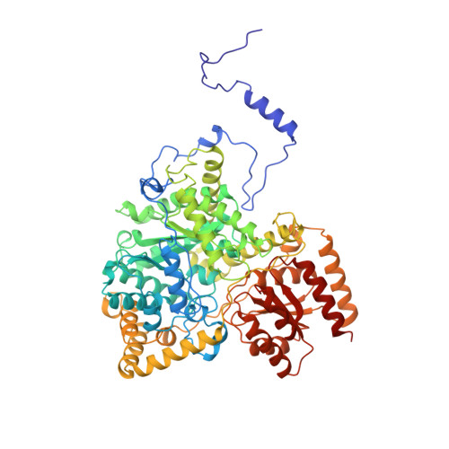
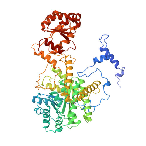
How coenzyme B12 radicals are generated: the crystal structure of methylmalonyl-coenzyme A mutase at 2 A resolution.

Mancia, F.Keep, N.H.Nakagawa, A.Leadlay, P.F.McSweeney, S.Rasmussen, B.Bosecke, P.Diat, O.Evans, P.R.

(1996) Structure 4: 339-350

  • DOI: https://doi.org/10.1016/s0969-2126(96)00037-8
  • Primary Citation of Related Structures:  
    1REQ

  • PubMed Abstract: 

    The enzyme methylmalonyl-coenzyme A (CoA) mutase, an alphabeta heterodimer of 150 kDa, is a member of a class of enzymes that uses coenzyme B12 (adenosylcobalamin) as a cofactor. The enzyme induces the formation of an adenosyl radical from the cofactor. This radical then initiates a free-radical rearrangement of its substrate, succinyl-CoA, to methylmalonyl-CoA. Reported here is the crystal structure at 2 A resolution of methylmalonyl-CoA mutase from Propionibacterium shermanii in complex with coenzyme B12 and with the partial substrate desulpho-CoA (lacking the succinyl group and the sulphur atom of the substrate). The coenzyme is bound by a domain which shares a similar fold to those of flavodoxin and the B12-binding domain of methylcobalamin-dependent methionine synthase. The cobalt atom is coordinated, via a long bond, to a histidine from the protein. The partial substrate is bound along the axis of a (beta/alpha)8 TIM barrel domain. The histidine-cobalt distance is very long (2.5 A compared with 1.95-2.2 A in free cobalamins), suggesting that the enzyme positions the histidine in order to weaken the metal-carbon bond of the cofactor and favour the formation of the initial radical species. The active site is deeply buried, and the only access to it is through a narrow tunnel along the axis of the TIM barrel domain.


  • Organizational Affiliation

    MRC Laboratory of Molecular Biology, Hills Road, Cambridge CB2 2QH, UK.


Macromolecules
Find similar proteins by:  (by identity cutoff)  |  3D Structure
Entity ID: 1
MoleculeChains Sequence LengthOrganismDetailsImage
METHYLMALONYL-COA MUTASE
A, C
727Propionibacterium freudenreichii subsp. shermaniiMutation(s): 0 
Gene Names: MUTA MUTB
EC: 5.4.99.2
UniProt
Find proteins for P11653 (Propionibacterium freudenreichii subsp. shermanii)
Explore P11653 
Go to UniProtKB:  P11653
Entity Groups  
Sequence Clusters30% Identity50% Identity70% Identity90% Identity95% Identity100% Identity
UniProt GroupP11653
Sequence Annotations
Expand
  • Reference Sequence
Find similar proteins by:  (by identity cutoff)  |  3D Structure
Entity ID: 2
MoleculeChains Sequence LengthOrganismDetailsImage
METHYLMALONYL-COA MUTASE
B, D
637Propionibacterium freudenreichii subsp. shermaniiMutation(s): 0 
Gene Names: MUTA MUTB
EC: 5.4.99.2
UniProt
Find proteins for P11652 (Propionibacterium freudenreichii subsp. shermanii)
Explore P11652 
Go to UniProtKB:  P11652
Entity Groups  
Sequence Clusters30% Identity50% Identity70% Identity90% Identity95% Identity100% Identity
UniProt GroupP11652
Sequence Annotations
Expand
  • Reference Sequence
Small Molecules
Ligands 3 Unique
IDChains Name / Formula / InChI Key2D Diagram3D Interactions
B12
Query on B12

Download Ideal Coordinates CCD File 
E [auth A],
L [auth C]
COBALAMIN
C62 H89 Co N13 O14 P
LKVIQTCSMMVGFU-DWSMJLPVSA-N
DCA
Query on DCA

Download Ideal Coordinates CCD File 
F [auth A],
M [auth C]
DESULFO-COENZYME A
C21 H36 N7 O16 P3
ILWZMFJBPIYQKW-IBOSZNHHSA-N
GOL
Query on GOL

Download Ideal Coordinates CCD File 
G [auth A]
H [auth A]
I [auth B]
J [auth B]
K [auth B]
G [auth A],
H [auth A],
I [auth B],
J [auth B],
K [auth B],
N [auth C],
O [auth C],
P [auth D],
Q [auth D]
GLYCEROL
C3 H8 O3
PEDCQBHIVMGVHV-UHFFFAOYSA-N
Experimental Data & Validation

Experimental Data

  • Method: X-RAY DIFFRACTION
  • Resolution: 2.00 Å
  • R-Value Free:  0.275 (Depositor), 0.270 (DCC) 
  • R-Value Work:  0.220 (Depositor), 0.250 (DCC) 
Space Group: P 1 21 1
Unit Cell:
Length ( Å )Angle ( ˚ )
a = 119.8α = 90
b = 161.3β = 105.1
c = 88.4γ = 90
Software Package:
Software NamePurpose
REFMACrefinement
MOSFLMdata reduction

Structure Validation

View Full Validation Report


Ligand Structure Quality Assessment 


Entry History 

Deposition Data

Revision History  (Full details and data files)

  • Version 1.0: 1997-01-27
    Type: Initial release
  • Version 1.1: 2008-03-24
    Changes: Version format compliance
  • Version 1.2: 2011-07-13
    Changes: Non-polymer description, Version format compliance
  • Version 1.3: 2012-10-24
    Changes: Non-polymer description
  • Version 1.4: 2024-02-14
    Changes: Data collection, Database references, Derived calculations, Other