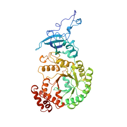
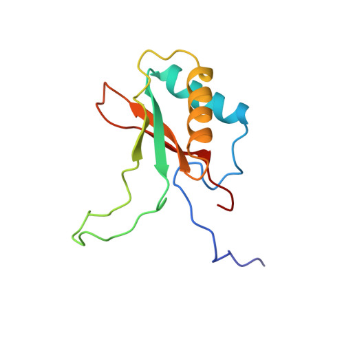
1RXO

ACTIVATED SPINACH RUBISCO IN COMPLEX WITH ITS SUBSTRATE RIBULOSE-1,5-BISPHOSPHATE AND CALCIUM


Experimental Data Snapshot

  • Method: X-RAY DIFFRACTION
  • Resolution: 2.20 Å
  • R-Value Free: 0.229 
  • R-Value Work: 0.205 
  • R-Value Observed: 0.205 

wwPDB Validation   3D Report Full Report


Ligand Structure Quality Assessment 


This is version 1.3 of the entry. See complete history


Literature

The structure of the complex between rubisco and its natural substrate ribulose 1,5-bisphosphate.

Taylor, T.C.Andersson, I.

(1997) J Mol Biol 265: 432-444

  • DOI: https://doi.org/10.1006/jmbi.1996.0738
  • Primary Citation of Related Structures:  
    1RCX, 1RXO

  • PubMed Abstract: 

    The three-dimensional structure of the complex of ribulose 1,5-bisphosphate carboxylase/oxygenase (rubisco; EC 4.1.1.39) from spinach with its natural substrate ribulose 1,5-bisphosphate (RuBP) has been determined both under activating and non-activating conditions by X-ray crystallography to a resolution of 2.1 A and 2.4 A, respectively. Under activating conditions, the use of calcium instead of magnesium as the activator metal ion enabled us to trap the substrate in a stable complex for crystallographic analysis. Comparison of the structure of the activated and the non-activated RuBP complexes shows a tighter binding for the substrate in the non-activated form of the enzyme, in line with previous solution studies. In the non-activated complex, the substrate triggers isolation of the active site by inducing movements of flexible loop regions of the catalytic subunits. In contrast, in the activated complex the active site remains partly open, probably awaiting the binding of the gaseous substrate. By inspection of the structures and by comparison with other complexes of the enzyme we were able to identify a network of hydrogen bonds that stabilise a closed active site structure during crucial steps in the reaction. The present structure underlines the central role of the carbamylated lysine 201 in both activation and catalysis, and completes available structural information for our proposal on the mechanism of the enzyme.


  • Organizational Affiliation

    Department of Molecular Biology, Swedish University of Agricultural Sciences, Uppsala.


Macromolecules
Find similar proteins by:  (by identity cutoff)  |  3D Structure
Entity ID: 1
MoleculeChains Sequence LengthOrganismDetailsImage
RIBULOSE BISPHOSPHATE CARBOXYLASE/OXYGENASEA [auth L],
C [auth B],
E,
G [auth H]
475Spinacia oleraceaMutation(s): 1 
EC: 4.1.1.39
UniProt
Find proteins for P00875 (Spinacia oleracea)
Explore P00875 
Go to UniProtKB:  P00875
Entity Groups  
Sequence Clusters30% Identity50% Identity70% Identity90% Identity95% Identity100% Identity
UniProt GroupP00875
Sequence Annotations
Expand
  • Reference Sequence
Find similar proteins by:  (by identity cutoff)  |  3D Structure
Entity ID: 2
MoleculeChains Sequence LengthOrganismDetailsImage
RIBULOSE BISPHOSPHATE CARBOXYLASE/OXYGENASEB [auth S],
D [auth C],
F,
H [auth I]
123Spinacia oleraceaMutation(s): 0 
EC: 4.1.1.39
UniProt
Find proteins for P00870 (Spinacia oleracea)
Explore P00870 
Go to UniProtKB:  P00870
Entity Groups  
Sequence Clusters30% Identity50% Identity70% Identity90% Identity95% Identity100% Identity
UniProt GroupP00870
Sequence Annotations
Expand
  • Reference Sequence
Small Molecules
Ligands 2 Unique
IDChains Name / Formula / InChI Key2D Diagram3D Interactions
RUB
Query on RUB

Download Ideal Coordinates CCD File 
I [auth L],
K [auth B],
M [auth E],
O [auth H]
RIBULOSE-1,5-DIPHOSPHATE
C5 H12 O11 P2
YAHZABJORDUQGO-NQXXGFSBSA-N
CA
Query on CA

Download Ideal Coordinates CCD File 
J [auth L],
L [auth B],
N [auth E],
P [auth H]
CALCIUM ION
Ca
BHPQYMZQTOCNFJ-UHFFFAOYSA-N
Modified Residues  1 Unique
IDChains TypeFormula2D DiagramParent
KCX
Query on KCX
A [auth L],
C [auth B],
E,
G [auth H]
L-PEPTIDE LINKINGC7 H14 N2 O4LYS
Experimental Data & Validation

Experimental Data

  • Method: X-RAY DIFFRACTION
  • Resolution: 2.20 Å
  • R-Value Free: 0.229 
  • R-Value Work: 0.205 
  • R-Value Observed: 0.205 
  • Space Group: C 2 2 21
Unit Cell:
Length ( Å )Angle ( ˚ )
a = 157.6α = 90
b = 158.5β = 90
c = 202.6γ = 90
Software Package:
Software NamePurpose
X-PLORmodel building
X-PLORrefinement
DENZOdata reduction
SCALEPACKdata scaling
X-PLORphasing

Structure Validation

View Full Validation Report


Ligand Structure Quality Assessment 


Entry History 

Deposition Data

Revision History  (Full details and data files)

  • Version 1.0: 1997-06-16
    Type: Initial release
  • Version 1.1: 2008-03-24
    Changes: Version format compliance
  • Version 1.2: 2011-07-13
    Changes: Derived calculations, Version format compliance
  • Version 1.3: 2024-06-05
    Changes: Data collection, Database references, Derived calculations, Other