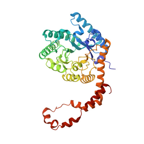
1S5N

Xylose Isomerase in Substrate and Inhibitor Michaelis States: Atomic Resolution Studies of a Metal-Mediated Hydride Shift


Experimental Data Snapshot

  • Method: X-RAY DIFFRACTION
  • Resolution: 0.95 Å
  • R-Value Free: 0.128 
  • R-Value Work: 0.106 

Starting Model: experimental
View more details

wwPDB Validation   3D Report Full Report


Ligand Structure Quality Assessment 


This is version 1.6 of the entry. See complete history


Literature

Xylose isomerase in substrate and inhibitor michaelis States: atomic resolution studies of a metal-mediated hydride shift(,).

Fenn, T.D.Ringe, D.Petsko, G.A.

(2004) Biochemistry 43: 6464-6474

  • DOI: https://doi.org/10.1021/bi049812o
  • Primary Citation of Related Structures:  
    1S5M, 1S5N

  • PubMed Abstract: 

    Xylose isomerase (E.C. 5.3.1.5) catalyzes the interconversion of aldose and ketose sugars and has an absolute requirement for two divalent cations at its active site to drive the hydride transfer rates of sugar isomerization. Evidence suggests some degree of metal movement at the second metal site, although how this movement may affect catalysis is unknown. The 0.95 A resolution structure of the xylitol-inhibited enzyme presented here suggests three alternative positions for the second metal ion, only one of which appears positioned in a catalytically competent manner. To complete the reaction, an active site hydroxyl species appears appropriately positioned for hydrogen transfer, as evidenced by precise bonding distances. Conversely, the 0.98 A resolution structure of the enzyme with glucose bound in the alpha-pyranose state only shows one of the metal ion conformations at the second metal ion binding site, suggesting that the linear form of the sugar is required to promote the second and third metal ion conformations. The two structures suggest a strong degree of conformational flexibility at the active site, which seems required for catalysis and may explain the poor rate of turnover for this enzyme. Further, the pyranose structure implies that His53 may act as the initial acid responsible for ring opening of the sugar to the aldose form, an observation that has been difficult to establish in previous studies. The glucose ring also appears to display significant segmented disorder in a manner suggestive of ring opening, perhaps lending insight into means of enzyme destabilization of the ground state to promote catalysis. On the basis of these results, we propose a modified version of the bridged bimetallic mechanism for hydride transfer in the case of Streptomyces olivochromogenes xylose isomerase.


  • Organizational Affiliation

    Department of Biochemistry, Graduate Program in Biophysics, Rosenstiel Basic Medical Sciences Research Center, Brandeis University, Waltham, Massachusetts 02454-9110, USA.


Macromolecules
Find similar proteins by:  (by identity cutoff)  |  3D Structure
Entity ID: 1
MoleculeChains Sequence LengthOrganismDetailsImage
Xylose isomerase386Streptomyces olivochromogenesMutation(s): 0 
Gene Names: XYLA
EC: 5.3.1.5
UniProt
Find proteins for P15587 (Streptomyces olivochromogenes)
Explore P15587 
Go to UniProtKB:  P15587
Entity Groups  
Sequence Clusters30% Identity50% Identity70% Identity90% Identity95% Identity100% Identity
UniProt GroupP15587
Sequence Annotations
Expand
  • Reference Sequence
Small Molecules
Ligands 4 Unique
IDChains Name / Formula / InChI Key2D Diagram3D Interactions
XYL
Query on XYL

Download Ideal Coordinates CCD File 
B [auth A]Xylitol
C5 H12 O5
HEBKCHPVOIAQTA-SCDXWVJYSA-N
MN
Query on MN

Download Ideal Coordinates CCD File 
C [auth A],
D [auth A]
MANGANESE (II) ION
Mn
WAEMQWOKJMHJLA-UHFFFAOYSA-N
NA
Query on NA

Download Ideal Coordinates CCD File 
E [auth A],
F [auth A]
SODIUM ION
Na
FKNQFGJONOIPTF-UHFFFAOYSA-N
OH
Query on OH

Download Ideal Coordinates CCD File 
G [auth A]HYDROXIDE ION
H O
XLYOFNOQVPJJNP-UHFFFAOYSA-M
Experimental Data & Validation

Experimental Data

  • Method: X-RAY DIFFRACTION
  • Resolution: 0.95 Å
  • R-Value Free: 0.128 
  • R-Value Work: 0.106 
  • Space Group: I 2 2 2
Unit Cell:
Length ( Å )Angle ( ˚ )
a = 86.031α = 90
b = 92.854β = 90
c = 98.312γ = 90
Software Package:
Software NamePurpose
SHELXL-97refinement
SCALEPACKdata scaling
CNSrefinement
HKL-2000data collection
HKL-2000data reduction
CNSphasing

Structure Validation

View Full Validation Report


Ligand Structure Quality Assessment 


Entry History 

Deposition Data

Revision History  (Full details and data files)

  • Version 1.0: 2004-02-10
    Type: Initial release
  • Version 1.1: 2008-04-29
    Changes: Version format compliance
  • Version 1.2: 2011-07-13
    Changes: Derived calculations, Version format compliance
  • Version 1.3: 2018-01-31
    Changes: Experimental preparation
  • Version 1.4: 2019-07-24
    Changes: Data collection, Refinement description
  • Version 1.5: 2020-07-29
    Type: Remediation
    Reason: Carbohydrate remediation
    Changes: Derived calculations, Structure summary
  • Version 1.6: 2023-08-23
    Changes: Data collection, Database references, Refinement description, Structure summary