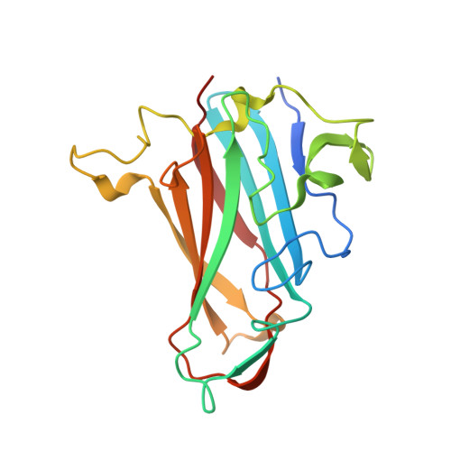
1UXB

ADENOVIRUS AD19p FIBRE HEAD in complex with sialyl-lactose


Experimental Data Snapshot

  • Method: X-RAY DIFFRACTION
  • Resolution: 1.75 Å
  • R-Value Free: 0.210 
  • R-Value Work: 0.175 
  • R-Value Observed: 0.175 

Starting Model: experimental
View more details

wwPDB Validation   3D Report Full Report


This is version 2.1 of the entry. See complete history


Literature

Crystal Structure of Species D Adenovirus Fiber Knobs and Their Sialic Acid Binding Sites

Burmeister, W.P.Guilligay, D.Cusack, S.Wadell, G.Arnberg, N.

(2004) J Virol 78: 7727

  • DOI: https://doi.org/10.1128/JVI.78.14.7727-7736.2004
  • Primary Citation of Related Structures:  
    1UXA, 1UXB, 1UXE

  • PubMed Abstract: 

    Adenovirus serotype 37 (Ad37) belongs to species D and can cause epidemic keratoconjunctivitis, whereas the closely related Ad19p does not. Primary cell attachment by adenoviruses is mediated through receptor binding of the knob domain of the fiber protein. The knobs of Ad37 and Ad19p differ at only two positions, Lys240Glu and Asn340Asp. We report the high-resolution crystal structures of the Ad37 and Ad19p knobs, both native and in complex with sialic acid, which has been proposed as a receptor for Ad37. Overall, the Ad37 and Ad19p knobs are very similar to previously reported knob structures, especially to that of Ad5, which binds the coxsackievirus-adenovirus receptor (CAR). Ad37 and Ad19p knobs are structurally identical with the exception of the changed side chains and are structurally most similar to CAR-binding knobs (e.g., that of Ad5) rather than non-CAR-binding knobs (e.g., that of Ad3). The two mutations in Ad19p result in a partial loss of the exceptionally high positive surface charge of the Ad37 knob but do not affect sialic acid binding. This site is located on the top of the trimer and binds both alpha(2,3) and alpha(2,6)-linked sialyl-lactose, although only the sialic acid residue makes direct contact. Amino acid alignment suggests that the sialic acid binding site is conserved in several species D serotypes. Our results show that the altered viral tropism and cell binding of Ad19p relative to those of Ad37 are not explained by a different binding ability toward sialyl-lactose.


  • Organizational Affiliation

    Laboratoire de Virologie Moléculaire et Structurale, Université Joseph Fourier, Faculté de Médecine de Grenoble, F-38042 Grenoble cedex 9, France.


Macromolecules
Find similar proteins by:  (by identity cutoff)  |  3D Structure
Entity ID: 1
MoleculeChains Sequence LengthOrganismDetailsImage
FIBER PROTEIN
A, B, C
194Human adenovirus 19Mutation(s): 4 
UniProt
Find proteins for Q64822 (Human adenovirus 19)
Explore Q64822 
Go to UniProtKB:  Q64822
Entity Groups  
Sequence Clusters30% Identity50% Identity70% Identity90% Identity95% Identity100% Identity
UniProt GroupQ64822
Sequence Annotations
Expand
  • Reference Sequence
Oligosaccharides

Help

Entity ID: 2
MoleculeChains Length2D Diagram Glycosylation3D Interactions
N-acetyl-alpha-neuraminic acid-(2-3)-beta-D-galactopyranose
D, E
2N/A
Glycosylation Resources
GlyTouCan:  G30207PZ
GlyCosmos:  G30207PZ
GlyGen:  G30207PZ
Entity ID: 3
MoleculeChains Length2D Diagram Glycosylation3D Interactions
N-acetyl-alpha-neuraminic acid-(2-6)-beta-D-galactopyranose
F
2N/A
Glycosylation Resources
GlyTouCan:  G63069TR
GlyCosmos:  G63069TR
GlyGen:  G63069TR
Experimental Data & Validation

Experimental Data

  • Method: X-RAY DIFFRACTION
  • Resolution: 1.75 Å
  • R-Value Free: 0.210 
  • R-Value Work: 0.175 
  • R-Value Observed: 0.175 
  • Space Group: P 1 21 1
Unit Cell:
Length ( Å )Angle ( ˚ )
a = 60.7α = 90
b = 68.7β = 94.9
c = 74.7γ = 90
Software Package:
Software NamePurpose
CNSrefinement
MOSFLMdata reduction
SCALAdata scaling

Structure Validation

View Full Validation Report


Entry History 

Deposition Data

Revision History  (Full details and data files)

  • Version 1.0: 2004-07-01
    Type: Initial release
  • Version 1.1: 2011-05-07
    Changes: Version format compliance
  • Version 1.2: 2011-07-13
    Changes: Version format compliance
  • Version 2.0: 2020-07-29
    Type: Remediation
    Reason: Carbohydrate remediation
    Changes: Advisory, Atomic model, Data collection, Derived calculations, Other, Structure summary
  • Version 2.1: 2023-12-13
    Changes: Data collection, Database references, Refinement description, Structure summary