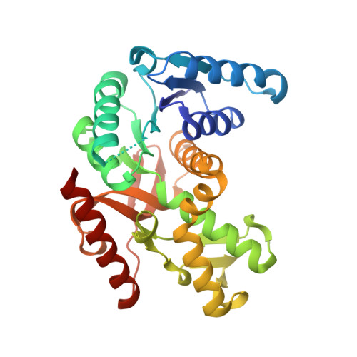
1UXK

Large improvement in the thermal stability of a tetrameric malate dehydrogenase by single point mutations at the dimer-dimer interface


Experimental Data Snapshot

  • Method: X-RAY DIFFRACTION
  • Resolution: 1.80 Å
  • R-Value Free: 0.214 
  • R-Value Work: 0.178 
  • R-Value Observed: 0.178 

Starting Model: experimental
View more details

wwPDB Validation   3D Report Full Report


Ligand Structure Quality Assessment 


This is version 1.4 of the entry. See complete history


Literature

Large Improvement in the Thermal Stability of a Tetrameric Malate Dehydrogenase by Single Point Mutations at the Dimer-Dimer Interface.

Bjork, A.Dalhus, B.Mantzilas, D.Sirevag, R.Eijsink, V.G.H.

(2004) J Mol Biol 341: 1215

  • DOI: https://doi.org/10.1016/j.jmb.2004.06.079
  • Primary Citation of Related Structures:  
    1UXG, 1UXH, 1UXI, 1UXJ, 1UXK

  • PubMed Abstract: 

    The stability of tetrameric malate dehydrogenase from the green phototrophic bacterium Chloroflexus aurantiacus (CaMDH) is at least in part determined by electrostatic interactions at the dimer-dimer interface. Since previous studies had indicated that the thermal stability of CaMDH becomes lower with increasing pH, attempts were made to increase the stability by removal of (excess) negative charge at the dimer-dimer interface. Mutation of Glu165 to Gln or Lys yielded a dramatic increase in thermal stability at pH 7.5 (+23.6 -- + 23.9 degrees C increase in apparent t(m)) and a more moderate increase at pH 4.4 (+4.6 -- + 5.4 degrees C). The drastically increased stability at neutral pH was achieved without forfeiture of catalytic performance at low temperatures. The crystal structures of the two mutants showed only minor structural changes close to the mutated residues, and indicated that the observed stability effects are solely due to subtle changes in the complex network of electrostatic interactions in the dimer-dimer interface. Both mutations reduced the concentration dependency of thermal stability, suggesting that the oligomeric structure had been reinforced. Interestingly, the two mutations had similar effects on stability, despite the charge difference between the introduced side-chains. Together with the loss of concentration dependency, this may indicate that both E165Q and E165K stabilize CaMDH to such an extent that disruption of the inter-dimer electrostatic network around residue 165 no longer limits kinetic thermal stability.


  • Organizational Affiliation

    Department of Molecular Biosciences, University of Oslo, P.O. Box 1041, Blindern, N-0316 Oslo, Norway. alexandra.bjork@bio.uio.no


Macromolecules
Find similar proteins by:  (by identity cutoff)  |  3D Structure
Entity ID: 1
MoleculeChains Sequence LengthOrganismDetailsImage
MALATE DEHYDROGENASEA,
B [auth C]
309Chloroflexus aurantiacusMutation(s): 1 
EC: 1.1.1.37
UniProt
Find proteins for P80040 (Chloroflexus aurantiacus (strain ATCC 29366 / DSM 635 / J-10-fl))
Explore P80040 
Go to UniProtKB:  P80040
Entity Groups  
Sequence Clusters30% Identity50% Identity70% Identity90% Identity95% Identity100% Identity
UniProt GroupP80040
Sequence Annotations
Expand
  • Reference Sequence
Small Molecules
Ligands 4 Unique
IDChains Name / Formula / InChI Key2D Diagram3D Interactions
NAD
Query on NAD

Download Ideal Coordinates CCD File 
C [auth A],
P [auth C]
NICOTINAMIDE-ADENINE-DINUCLEOTIDE
C21 H27 N7 O14 P2
BAWFJGJZGIEFAR-NNYOXOHSSA-N
CD
Query on CD

Download Ideal Coordinates CCD File 
D [auth A]
E [auth A]
F [auth A]
G [auth A]
H [auth A]
D [auth A],
E [auth A],
F [auth A],
G [auth A],
H [auth A],
M [auth A],
Q [auth C],
R [auth C],
T [auth C],
U [auth C]
CADMIUM ION
Cd
WLZRMCYVCSSEQC-UHFFFAOYSA-N
CL
Query on CL

Download Ideal Coordinates CCD File 
I [auth A],
S [auth C]
CHLORIDE ION
Cl
VEXZGXHMUGYJMC-UHFFFAOYSA-M
NA
Query on NA

Download Ideal Coordinates CCD File 
J [auth A]
K [auth A]
L [auth A]
N [auth A]
O [auth A]
J [auth A],
K [auth A],
L [auth A],
N [auth A],
O [auth A],
V [auth C]
SODIUM ION
Na
FKNQFGJONOIPTF-UHFFFAOYSA-N
Experimental Data & Validation

Experimental Data

  • Method: X-RAY DIFFRACTION
  • Resolution: 1.80 Å
  • R-Value Free: 0.214 
  • R-Value Work: 0.178 
  • R-Value Observed: 0.178 
  • Space Group: P 31 2 1
Unit Cell:
Length ( Å )Angle ( ˚ )
a = 105.405α = 90
b = 105.405β = 90
c = 102.305γ = 120
Software Package:
Software NamePurpose
CNSrefinement
DENZOdata reduction
SCALEPACKdata scaling
CNSphasing

Structure Validation

View Full Validation Report


Ligand Structure Quality Assessment 


Entry History 

Deposition Data

Revision History  (Full details and data files)

  • Version 1.0: 2004-08-26
    Type: Initial release
  • Version 1.1: 2015-04-15
    Changes: Database references, Derived calculations, Non-polymer description, Other, Refinement description, Version format compliance
  • Version 1.2: 2019-05-22
    Changes: Data collection, Other, Refinement description
  • Version 1.3: 2019-07-24
    Changes: Data collection
  • Version 1.4: 2023-12-13
    Changes: Data collection, Database references, Derived calculations, Other, Refinement description