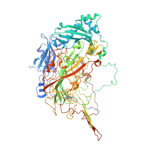
1W7C

PPLO at 1.23 Angstroms


Experimental Data Snapshot

  • Method: X-RAY DIFFRACTION
  • Resolution: 1.23 Å
  • R-Value Free: 0.146 
  • R-Value Work: 0.112 
  • R-Value Observed: 0.113 

Starting Model: experimental
View more details

wwPDB Validation   3D Report Full Report


Ligand Structure Quality Assessment 


This is version 2.1 of the entry. See complete history


Literature

The 1.23 A Structure of Pichia Pastoris Lysyl Oxidase Reveals a Lysine-Lysine Cross-Link

Duff, A.P.Cohen, A.E.Ellis, P.J.Hilmer, K.Langley, D.B.Dooley, D.M.Freeman, H.C.Guss, J.M.

(2006) Acta Crystallogr D Biol Crystallogr 62: 1073

  • DOI: https://doi.org/10.1107/S0907444906026333
  • Primary Citation of Related Structures:  
    1W7C

  • PubMed Abstract: 

    The structure of Pichia pastoris lysyl oxidase (PPLO) in a new crystal form has been refined at 1.23 Angstrom resolution. PPLO, a copper amine oxidase (CuAO) with a 2,4,5-trihydroxyphenylalanine quinone (TPQ) cofactor, differs from most other members of the CuAO enzyme family in having the ability to oxidize the side chain of lysine residues in a polypeptide. In the asymmetric unit of the crystals, the structure analysis has located residues 43-779 of the polypeptide chain, seven carbohydrate residues, the active-site Cu atom, an imidazole molecule bound at the active site, two buried Ca(2+) ions, five surface Mg(2+) ions, five surface Cl(-) ions and 1045 water molecules. The crystallographic residuals are R = 0.112 and R(free) = 0.146. The TPQ cofactor and several other active-site residues are poorly ordered, in contrast to the surrounding well ordered structure. A covalent cross-link is observed between two lysine residues, Lys778 and Lys66. The cross-link is likely to have been formed by the oxidation of Lys778 followed by a spontaneous reaction with Lys66. The link is modelled as dehydrolysinonorleucine.


  • Organizational Affiliation

    School of Molecular and Microbial Biosciences, University of Sydney, NSW 2006, Australia.


Macromolecules
Find similar proteins by:  (by identity cutoff)  |  3D Structure
Entity ID: 1
MoleculeChains Sequence LengthOrganismDetailsImage
LYSYL OXIDASE747Komagataella pastorisMutation(s): 0 
EC: 1.4.3.13 (PDB Primary Data), 1.4.3 (UniProt)
UniProt
Find proteins for Q96X16 (Komagataella pastoris)
Explore Q96X16 
Go to UniProtKB:  Q96X16
Entity Groups  
Sequence Clusters30% Identity50% Identity70% Identity90% Identity95% Identity100% Identity
UniProt GroupQ96X16
Glycosylation
Glycosylation Sites: 5
Sequence Annotations
Expand
  • Reference Sequence
Oligosaccharides

Help

Entity ID: 2
MoleculeChains Length2D Diagram Glycosylation3D Interactions
beta-D-mannopyranose-(1-4)-2-acetamido-2-deoxy-beta-D-glucopyranose-(1-4)-2-acetamido-2-deoxy-beta-D-glucopyranose
B
3N-Glycosylation
Glycosylation Resources
GlyTouCan:  G15407YE
GlyCosmos:  G15407YE
GlyGen:  G15407YE
Small Molecules
Ligands 6 Unique
IDChains Name / Formula / InChI Key2D Diagram3D Interactions
NAG
Query on NAG

Download Ideal Coordinates CCD File 
Q [auth A],
R [auth A],
S [auth A],
T [auth A]
2-acetamido-2-deoxy-beta-D-glucopyranose
C8 H15 N O6
OVRNDRQMDRJTHS-FMDGEEDCSA-N
IMD
Query on IMD

Download Ideal Coordinates CCD File 
P [auth A]IMIDAZOLE
C3 H5 N2
RAXXELZNTBOGNW-UHFFFAOYSA-O
CU
Query on CU

Download Ideal Coordinates CCD File 
C [auth A]COPPER (II) ION
Cu
JPVYNHNXODAKFH-UHFFFAOYSA-N
CA
Query on CA

Download Ideal Coordinates CCD File 
D [auth A],
E [auth A]
CALCIUM ION
Ca
BHPQYMZQTOCNFJ-UHFFFAOYSA-N
CL
Query on CL

Download Ideal Coordinates CCD File 
K [auth A],
L [auth A],
M [auth A],
N [auth A],
O [auth A]
CHLORIDE ION
Cl
VEXZGXHMUGYJMC-UHFFFAOYSA-M
MG
Query on MG

Download Ideal Coordinates CCD File 
F [auth A],
G [auth A],
H [auth A],
I [auth A],
J [auth A]
MAGNESIUM ION
Mg
JLVVSXFLKOJNIY-UHFFFAOYSA-N
Modified Residues  1 Unique
IDChains TypeFormula2D DiagramParent
TPQ
Query on TPQ
A
L-PEPTIDE LINKINGC9 H9 N O5TYR
Experimental Data & Validation

Experimental Data

  • Method: X-RAY DIFFRACTION
  • Resolution: 1.23 Å
  • R-Value Free: 0.146 
  • R-Value Work: 0.112 
  • R-Value Observed: 0.113 
  • Space Group: C 1 2 1
Unit Cell:
Length ( Å )Angle ( ˚ )
a = 139.948α = 90
b = 67.027β = 118.95
c = 108.688γ = 90
Software Package:
Software NamePurpose
REFMACrefinement
MOSFLMdata reduction
SCALAdata scaling
MOLREPphasing

Structure Validation

View Full Validation Report


Ligand Structure Quality Assessment 


Entry History 

Deposition Data

Revision History  (Full details and data files)

  • Version 1.0: 2006-08-31
    Type: Initial release
  • Version 1.1: 2013-01-16
    Changes: Atomic model, Database references, Derived calculations, Non-polymer description, Other, Refinement description, Source and taxonomy, Structure summary, Version format compliance
  • Version 1.2: 2019-01-30
    Changes: Advisory, Data collection, Experimental preparation, Other
  • Version 1.3: 2019-10-30
    Changes: Data collection, Derived calculations, Other
  • Version 2.0: 2020-07-29
    Type: Remediation
    Reason: Carbohydrate remediation
    Changes: Atomic model, Data collection, Derived calculations, Structure summary
  • Version 2.1: 2023-12-13
    Changes: Advisory, Data collection, Database references, Refinement description, Structure summary