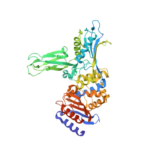
1W8Y

Crystal structure of the nitrocefin acyl-DD-peptidase from Actinomadura R39.


Experimental Data Snapshot

  • Method: X-RAY DIFFRACTION
  • Resolution: 2.40 Å
  • R-Value Free: 0.277 
  • R-Value Work: 0.220 
  • R-Value Observed: 0.220 

Starting Model: experimental
View more details

wwPDB Validation   3D Report Full Report


Ligand Structure Quality Assessment 


This is version 2.2 of the entry. See complete history


Literature

Crystal Structure of the Actinomadura R39 Dd- Peptidase Reveals New Domains in Penicillin- Binding Proteins.

Sauvage, E.Herman, R.Petrella, S.Duez, C.Bouillenne, F.Frere, J.M.Charlier, P.

(2005) J Biol Chem 280: 31249

  • DOI: https://doi.org/10.1074/jbc.M503271200
  • Primary Citation of Related Structures:  
    1W79, 1W8Q, 1W8Y

  • PubMed Abstract: 

    Actinomadura sp. R39 produces an exocellular DD-peptidase/penicillin-binding protein (PBP) whose primary structure is similar to that of Escherichia coli PBP4. It is characterized by a high beta-lactam-binding activity (second order rate constant for the acylation of the active site serine by benzylpenicillin: k2/K = 300 mm(-1) s(-1)). The crystal structure of the DD-peptidase from Actinomadura R39 was solved at a resolution of 1.8 angstroms by single anomalous dispersion at the cobalt resonance wavelength. The structure is composed of three domains: a penicillin-binding domain similar to the penicillin-binding domain of E. coli PBP5 and two domains of unknown function. In most multimodular PBPs, additional domains are generally located at the C or N termini of the penicillin-binding domain. In R39, the other two domains are inserted in the penicillin-binding domain, between the SXXK and SXN motifs, in a manner similar to "Matryoshka dolls." One of these domains is composed of a five-stranded beta-sheet with two helices on one side, and the other domain is a double three-stranded beta-sheet inserted in the previous domain. Additionally, the 2.4-angstroms structure of the acyl-enzyme complex of R39 with nitrocefin reveals the absence of active site conformational change upon binding the beta-lactams.


  • Organizational Affiliation

    Centre d'Ingénierie des Protéines, Université de Liège, Institut de Physique B5, B-4000 Liège, Belgium. eric.sauvage@ulg.ac.be


Macromolecules
Find similar proteins by:  (by identity cutoff)  |  3D Structure
Entity ID: 1
MoleculeChains Sequence LengthOrganismDetailsImage
D-alanyl-D-alanine carboxypeptidase
A, B, C, D
489Actinomadura sp. R39Mutation(s): 0 
EC: 3.4.16.4
UniProt
Find proteins for P39045 (Actinomadura sp. (strain R39))
Explore P39045 
Go to UniProtKB:  P39045
Entity Groups  
Sequence Clusters30% Identity50% Identity70% Identity90% Identity95% Identity100% Identity
UniProt GroupP39045
Sequence Annotations
Expand
  • Reference Sequence
Small Molecules
Ligands 3 Unique
IDChains Name / Formula / InChI Key2D Diagram3D Interactions
NCF
Query on NCF

Download Ideal Coordinates CCD File 
BA [auth D],
K [auth A],
P [auth B],
U [auth C]
(2R)-2-{(1R)-2-OXO-1-[(2-THIENYLACETYL)AMINO]ETHYL}-5,6-DIHYDRO-2H-1,3-THIAZINE-4-CARBOXYLIC ACID
C13 H14 N2 O5 S2
LNPNJPUNJYVKHH-WDEREUQCSA-N
SO4
Query on SO4

Download Ideal Coordinates CCD File 
E [auth A]
F [auth A]
G [auth A]
H [auth A]
L [auth B]
E [auth A],
F [auth A],
G [auth A],
H [auth A],
L [auth B],
M [auth B],
N [auth B],
O [auth B],
Q [auth C],
R [auth C],
S [auth C],
T [auth C],
V [auth D],
W [auth D],
X [auth D],
Y [auth D]
SULFATE ION
O4 S
QAOWNCQODCNURD-UHFFFAOYSA-L
MG
Query on MG

Download Ideal Coordinates CCD File 
AA [auth D],
I [auth A],
J [auth A],
Z [auth D]
MAGNESIUM ION
Mg
JLVVSXFLKOJNIY-UHFFFAOYSA-N
Experimental Data & Validation

Experimental Data

  • Method: X-RAY DIFFRACTION
  • Resolution: 2.40 Å
  • R-Value Free: 0.277 
  • R-Value Work: 0.220 
  • R-Value Observed: 0.220 
  • Space Group: P 1 21 1
Unit Cell:
Length ( Å )Angle ( ˚ )
a = 103.49α = 90
b = 94.36β = 94.58
c = 107.2γ = 90
Software Package:
Software NamePurpose
CNSrefinement
MOSFLMdata reduction
SCALAdata scaling

Structure Validation

View Full Validation Report


Ligand Structure Quality Assessment 


Entry History 

Deposition Data

Revision History  (Full details and data files)

  • Version 1.0: 2005-06-28
    Type: Initial release
  • Version 1.1: 2011-07-13
    Changes: Source and taxonomy, Version format compliance
  • Version 2.0: 2021-09-15
    Changes: Atomic model, Database references, Derived calculations, Non-polymer description, Other, Source and taxonomy, Structure summary
  • Version 2.1: 2023-12-13
    Changes: Data collection, Refinement description
  • Version 2.2: 2024-11-06
    Changes: Structure summary