1WZG | pdb_00001wzg

Crystal Structure Of An Artificial Metalloprotein: Fe(Salophen)/Wild Type Heme oxygenase


Experimental Data Snapshot

  • Method: X-RAY DIFFRACTION
  • Resolution: 1.75 Å
  • R-Value Free: 
    0.215 (Depositor), 0.207 (DCC) 
  • R-Value Work: 
    0.171 (Depositor) 
  • R-Value Observed: 
    0.176 (Depositor) 

wwPDB Validation 3D Report Full Report

Validation slider image for 1WZG

Ligand Structure Quality Assessment 


This is version 1.3 of the entry. See complete history

Literature

Design of metal cofactors activated by a protein-protein electron transfer system.

Ueno, T.Yokoi, N.Unno, M.Matsui, T.Tokita, Y.Yamada, M.Ikeda-Saito, M.Nakajima, H.Watanabe, Y.

(2006) Proc Natl Acad Sci U S A 103: 9416-9421

  • DOI: https://doi.org/10.1073/pnas.0510968103
  • Primary Citation Related Structures: 
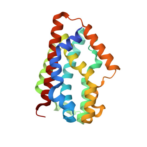
    1WZD, 1WZF, 1WZG

  • PubMed Abstract: 

    Protein-to-protein electron transfer (ET) is a critical process in biological chemistry for which fundamental understanding is expected to provide a wealth of applications in biotechnology. Investigations of protein-protein ET systems in reductive activation of artificial cofactors introduced into proteins remains particularly challenging because of the complexity of interactions between the cofactor and the system contributing to ET. In this work, we construct an artificial protein-protein ET system, using heme oxygenase (HO), which is known to catalyze the conversion of heme to biliverdin. HO uses electrons provided from NADPH/cytochrome P450 reductase (CPR) through protein-protein complex formation during the enzymatic reaction. We report that a Fe(III)(Schiff-base), in the place of the active-site heme prosthetic group of HO, can be reduced by NADPH/CPR. The crystal structure of the Fe(10-CH(2)CH(2)COOH-Schiff-base).HO composite indicates the presence of a hydrogen bond between the propionic acid carboxyl group and Arg-177 of HO. Furthermore, the ET rate from NADPH/CPR to the composite is 3.5-fold faster than that of Fe(Schiff-base).HO, although the redox potential of Fe(10-CH(2)CH(2)COOH-Schiff-base).HO (-79 mV vs. NHE) is lower than that of Fe(Schiff-base).HO (+15 mV vs. NHE), where NHE is normal hydrogen electrode. This work describes a synthetic metal complex activated by means of a protein-protein ET system, which has not previously been reported. Moreover, the result suggests the importance of the hydrogen bond for the ET reaction of HO. Our Fe(Schiff-base).HO composite model system may provide insights with regard to design of ET biosystems for sensors, catalysts, and electronics devices.


  • Organizational Affiliation
    • Research Center for Materials Science, Graduate School of Science, Nagoya University, Furo-cho, Chikusa-ku, Nagoya 464-8602, Japan.

Macromolecule Content 

  • Total Structure Weight: 49.75 kDa 
  • Atom Count: 3,763 
  • Modeled Residue Count: 418 
  • Deposited Residue Count: 430 
  • Unique protein chains: 1

Macromolecules

Find similar proteins by:|  3D Structure
Entity ID: 1
MoleculeChains  Sequence LengthOrganismDetailsImage
Heme oxygenase
A, B
215Corynebacterium diphtheriaeMutation(s): 0 
EC: 1.14.99.3 (PDB Primary Data), 1.14.14.18 (UniProt)
UniProt
Find proteins for P71119 (Corynebacterium diphtheriae (strain ATCC 700971 / NCTC 13129 / Biotype gravis))
Explore P71119 
Go to UniProtKB:  P71119
Entity Groups
Sequence Clusters30% Identity50% Identity70% Identity90% Identity95% Identity100% Identity
UniProt GroupP71119
Sequence Annotations
Expand
Reference Sequence

Small Molecules

Ligands 3 Unique
IDChains Name / Formula / InChI Key2D Diagram3D Interactions
YOM

Query on YOM


Download:Ideal Coordinates CCD File
H [auth A],
J [auth B]
2,2'-[1,2-PHENYLENEBIS(NITRILOMETHYLIDYNE)]BIS[PHENOLATO]](2-)-N,N',O,O']-IRON
C20 H14 Fe N2 O2
BSPYTUHYTMXMCB-JKBLJYNNSA-L
SO4

Query on SO4


Download:Ideal Coordinates CCD File
C [auth A]
D [auth A]
E [auth A]
F [auth A]
G [auth A]
C [auth A],
D [auth A],
E [auth A],
F [auth A],
G [auth A],
I [auth B]
SULFATE ION
O4 S
QAOWNCQODCNURD-UHFFFAOYSA-L
GOL

Query on GOL


Download:Ideal Coordinates CCD File
K [auth B]GLYCEROL
C3 H8 O3
PEDCQBHIVMGVHV-UHFFFAOYSA-N

Experimental Data & Validation

Experimental Data

  • Method: X-RAY DIFFRACTION
  • Resolution: 1.75 Å
  • R-Value Free:  0.215 (Depositor), 0.207 (DCC) 
  • R-Value Work:  0.171 (Depositor) 
  • R-Value Observed: 0.176 (Depositor) 
Space Group: P 1 21 1
Unit Cell:
Length ( Å )Angle ( ˚ )
a = 41.471α = 90
b = 62.884β = 98.89
c = 77.991γ = 90
Software Package:
Software NamePurpose
REFMACrefinement
HKL-2000data reduction
SCALEPACKdata scaling
CNSphasing

Structure Validation

View Full Validation Report


Ligand Structure Quality Assessment 


Entry History 

Deposition Data

Revision History  (Full details and data files)

  • Version 1.0: 2006-02-21
    Type: Initial release
  • Version 1.1: 2008-04-30
    Changes: Version format compliance
  • Version 1.2: 2011-07-13
    Changes: Non-polymer description, Version format compliance
  • Version 1.3: 2024-03-13
    Changes: Data collection, Database references, Derived calculations