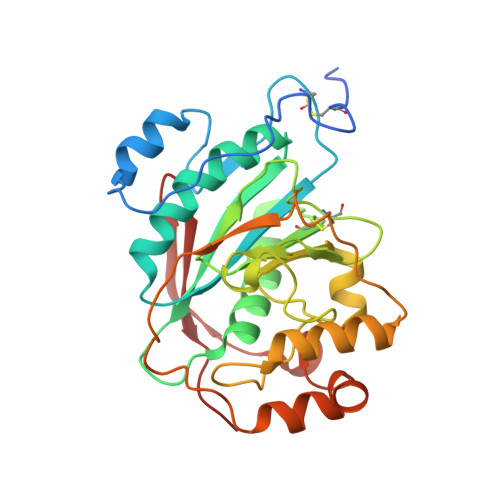
2AGD

Crystal Structure of Human M340H-Beta-1,4-Galactosyltransferase-I(M340H-B4Gal-T1) in Complex with GlcNAc-beta1,4-Man-alpha1,3-Man-beta-OR


Experimental Data Snapshot

  • Method: X-RAY DIFFRACTION
  • Resolution: 1.90 Å
  • R-Value Free: 0.228 
  • R-Value Work: 0.202 
  • R-Value Observed: 0.202 

Starting Model: experimental
View more details

wwPDB Validation   3D Report Full Report


Ligand Structure Quality Assessment 


This is version 2.3 of the entry. See complete history


Literature

Oligosaccharide Preferences of beta1,4-Galactosyltransferase-I: Crystal Structures of Met340His Mutant of Human beta1,4-Galactosyltransferase-I with a Pentasaccharide and Trisaccharides of the N-Glycan Moiety

Ramasamy, V.Ramakrishnan, B.Boeggeman, E.Ratner, D.M.Seeberger, P.H.Qasba, P.K.

(2005) J Mol Biol 353: 53-67

  • DOI: https://doi.org/10.1016/j.jmb.2005.07.050
  • Primary Citation of Related Structures:  
    2AE7, 2AEC, 2AES, 2AGD, 2AH9

  • PubMed Abstract: 

    beta-1,4-Galactosyltransferase-I (beta4Gal-T1) transfers galactose from UDP-galactose to N-acetylglucosamine (GlcNAc) residues of the branched N-linked oligosaccharide chains of glycoproteins. In an N-linked biantennary oligosaccharide chain, one antenna is attached to the 3-hydroxyl-(1,3-arm), and the other to the 6-hydroxyl-(1,6-arm) group of mannose, which is beta-1,4-linked to an N-linked chitobiose, attached to the aspargine residue of a protein. For a better understanding of the branch specificity of beta4Gal-T1 towards the GlcNAc residues of N-glycans, we have carried out kinetic and crystallographic studies with the wild-type human beta4Gal-T1 (h-beta4Gal-T1) and the mutant Met340His-beta4Gal-T1 (h-M340H-beta4Gal-T1) in complex with a GlcNAc-containing pentasaccharide and several GlcNAc-containing trisaccharides present in N-glycans. The oligosaccharides used were: pentasaccharide GlcNAcbeta1,2-Manalpha1,6 (GlcNAcbeta1,2-Manalpha1,3)Man; the 1,6-arm trisaccharide, GlcNAcbeta1,2-Manalpha1,6-Manbeta-OR (1,2-1,6-arm); the 1,3-arm trisaccharides, GlcNAcbeta1,2-Manalpha1,3-Manbeta-OR (1,2-1,3-arm) and GlcNAcbeta1,4-Manalpha1,3-Manbeta-OR (1,4-1,3-arm); and the trisaccharide GlcNAcbeta1,4-GlcNAcbeta1,4-GlcNAc (chitotriose). With the wild-type h-beta4Gal-T1, the K(m) of 1,2-1,6-arm is approximately tenfold lower than for 1,2-1,3-arm and 1,4-1,3-arm, and 22-fold lower than for chitotriose. Crystal structures of h-M340H-beta4Gal-T1 in complex with the pentasaccharide and various trisaccharides at 1.9-2.0A resolution showed that beta4Gal-T1 is in a closed conformation with the oligosaccharide bound to the enzyme, and the 1,2-1,6-arm trisaccharide makes the maximum number of interactions with the enzyme, which is in concurrence with the lowest K(m) for the trisaccharide. Present studies suggest that beta4Gal-T1 interacts preferentially with the 1,2-1,6-arm trisaccharide rather than with the 1,2-1,3-arm or 1,4-1,3-arm of a bi- or tri-antennary oligosaccharide chain of N-glycan.


  • Organizational Affiliation

    Laboratory of Experimental and Computational Biology, Center for Cancer Research, National Cancer Institute at Frederick, Frederick, MD 21702, USA.


Macromolecules
Find similar proteins by:  (by identity cutoff)  |  3D Structure
Entity ID: 1
MoleculeChains Sequence LengthOrganismDetailsImage
Beta-1,4-galactosyltransferase 1
A, B, C
287Homo sapiensMutation(s): 3 
Gene Names: B4GALT1GGTB2
EC: 2.4.1.90 (PDB Primary Data), 2.4.1 (UniProt), 2.4.1.22 (UniProt), 2.4.1.275 (UniProt), 2.4.1.38 (UniProt)
UniProt & NIH Common Fund Data Resources
Find proteins for P15291 (Homo sapiens)
Explore P15291 
Go to UniProtKB:  P15291
PHAROS:  P15291
GTEx:  ENSG00000086062 
Entity Groups  
Sequence Clusters30% Identity50% Identity70% Identity90% Identity95% Identity100% Identity
UniProt GroupP15291
Sequence Annotations
Expand
  • Reference Sequence
Oligosaccharides

Help

Entity ID: 2
MoleculeChains Length2D Diagram Glycosylation3D Interactions
2-acetamido-2-deoxy-beta-D-glucopyranose-(1-4)-alpha-D-mannopyranose-(1-3)-beta-D-mannopyranose
D, E, F
3N/A
Glycosylation Resources
GlyTouCan:  G98135ER
GlyCosmos:  G98135ER
GlyGen:  G98135ER
Small Molecules
Ligands 6 Unique
IDChains Name / Formula / InChI Key2D Diagram3D Interactions
UDH
Query on UDH

Download Ideal Coordinates CCD File 
IA [auth C],
L [auth A],
X [auth B]
6-AMINOHEXYL-URIDINE-C1,5'-DIPHOSPHATE
C15 H27 N3 O12 P2
MLWJBKPFDKRHBM-FMKGYKFTSA-N
MES
Query on MES

Download Ideal Coordinates CCD File 
KA [auth C]2-(N-MORPHOLINO)-ETHANESULFONIC ACID
C6 H13 N O4 S
SXGZJKUKBWWHRA-UHFFFAOYSA-N
SO4
Query on SO4

Download Ideal Coordinates CCD File 
DA [auth C]
EA [auth C]
FA [auth C]
GA [auth C]
H [auth A]
DA [auth C],
EA [auth C],
FA [auth C],
GA [auth C],
H [auth A],
HA [auth C],
I [auth A],
J [auth A],
K [auth A],
Q [auth B],
R [auth B],
S [auth B],
T [auth B],
U [auth B],
V [auth B],
W [auth B]
SULFATE ION
O4 S
QAOWNCQODCNURD-UHFFFAOYSA-L
GOL
Query on GOL

Download Ideal Coordinates CCD File 
AA [auth B]
BA [auth B]
LA [auth C]
M [auth A]
N [auth A]
AA [auth B],
BA [auth B],
LA [auth C],
M [auth A],
N [auth A],
O [auth A],
Y [auth B],
Z [auth B]
GLYCEROL
C3 H8 O3
PEDCQBHIVMGVHV-UHFFFAOYSA-N
DIO
Query on DIO

Download Ideal Coordinates CCD File 
JA [auth C]1,4-DIETHYLENE DIOXIDE
C4 H8 O2
RYHBNJHYFVUHQT-UHFFFAOYSA-N
MN
Query on MN

Download Ideal Coordinates CCD File 
CA [auth C],
G [auth A],
P [auth B]
MANGANESE (II) ION
Mn
WAEMQWOKJMHJLA-UHFFFAOYSA-N
Experimental Data & Validation

Experimental Data

  • Method: X-RAY DIFFRACTION
  • Resolution: 1.90 Å
  • R-Value Free: 0.228 
  • R-Value Work: 0.202 
  • R-Value Observed: 0.202 
  • Space Group: C 2 2 21
Unit Cell:
Length ( Å )Angle ( ˚ )
a = 107.464α = 90
b = 194.784β = 90
c = 143.7γ = 90
Software Package:
Software NamePurpose
HKL-2000data collection
SCALEPACKdata scaling
AMoREphasing
CNSrefinement
HKL-2000data reduction

Structure Validation

View Full Validation Report


Ligand Structure Quality Assessment 


Entry History 

Deposition Data

Revision History  (Full details and data files)

  • Version 1.0: 2005-10-04
    Type: Initial release
  • Version 1.1: 2008-04-30
    Changes: Version format compliance
  • Version 1.2: 2011-07-13
    Changes: Non-polymer description, Version format compliance
  • Version 2.0: 2020-07-29
    Type: Remediation
    Reason: Carbohydrate remediation
    Changes: Atomic model, Data collection, Derived calculations, Structure summary
  • Version 2.1: 2021-10-20
    Changes: Database references, Structure summary
  • Version 2.2: 2023-08-23
    Changes: Data collection, Refinement description
  • Version 2.3: 2024-10-16
    Changes: Structure summary