2AP2 | pdb_00002ap2

SINGLE CHAIN FV OF C219 IN COMPLEX WITH SYNTHETIC EPITOPE PEPTIDE


Experimental Data Snapshot

  • Method: X-RAY DIFFRACTION
  • Resolution: 2.40 Å
  • R-Value Free: 
    0.274 (Depositor), 0.267 (DCC) 
  • R-Value Work: 
    0.221 (Depositor), 0.223 (DCC) 

Starting Model: experimental
View more details

wwPDB Validation 3D Report Full Report

Validation slider image for 2AP2

This is version 2.3 of the entry. See complete history

Literature

Antibody C219 recognizes an alpha-helical epitope on P-glycoprotein.

van Den Elsen, J.M.Kuntz, D.A.Hoedemaeker, F.J.Rose, D.R.

(1999) Proc Natl Acad Sci U S A 96: 13679-13684

  • DOI: https://doi.org/10.1073/pnas.96.24.13679
  • Primary Citation Related Structures: 
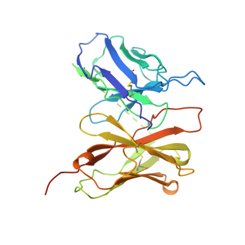
    2AP2

  • PubMed Abstract: 

    The ABC transporter, P-glycoprotein, is an integral membrane protein that mediates the ATP-driven efflux of drugs from multidrug-resistant cancer and HIV-infected cells. Anti-P-glycoprotein antibody C219 binds to both of the ATP-binding regions of P-glycoprotein and has been shown to inhibit its ATPase activity and drug binding capacity. C219 has been widely used in a clinical setting as a tumor marker, but recent observations of cross-reactivity with other proteins, including the c-erbB2 protein in breast cancer cells, impose potential limitations in detecting P-glycoprotein. We have determined the crystal structure at a resolution of 2.4 A of the variable fragment of C219 in complex with an epitope peptide derived from the nucleotide binding domain of P-glycoprotein. The 14-residue peptide adopts an amphipathic alpha-helical conformation, a secondary structure not previously observed in structures of antibody-peptide complexes. Together with available biochemical data, the crystal structure of the C219-peptide complex indicates the molecular basis of the cross-reactivity of C219 with non-multidrug resistance-associated proteins. Alignment of the C219 epitope with the recent crystal structure of the ATP-binding subunit of histidine permease suggests a structural basis for the inhibition of the ATP and drug binding capacity of P-glycoprotein by C219. The results provide a rationale for the development of C219 mutants with improved specificity and affinity that could be useful in antibody-based P-glycoprotein detection and therapy in multidrug resistant cancers.


  • Organizational Affiliation
    • Ontario Cancer Institute, Department of Medical Biophysics, University of Toronto, 610 University Avenue, Toronto M5G 2M9, Ontario, Canada.

Macromolecule Content 

  • Total Structure Weight: 61.79 kDa 
  • Atom Count: 3,954 
  • Modeled Residue Count: 506 
  • Deposited Residue Count: 570 
  • Unique protein chains: 2

Macromolecules

Find similar proteins by:|  3D Structure
Entity ID: 1
MoleculeChains  Sequence LengthOrganismDetailsImage
SINGLE CHAIN FVA,
B [auth C]
271Mus musculusMutation(s): 0 
UniProt
Find proteins for Q505N9 (Mus musculus)
Explore Q505N9 
Go to UniProtKB:  Q505N9
Find proteins for Q6KB05 (Mus musculus)
Explore Q6KB05 
Go to UniProtKB:  Q6KB05
Entity Groups
Sequence Clusters30% Identity50% Identity70% Identity90% Identity95% Identity100% Identity
UniProt GroupsQ505N9Q6KB05
Sequence Annotations
Expand
Reference Sequence
Find similar proteins by:  Sequence   |   3D Structure  
Entity ID: 2
MoleculeChains  Sequence LengthOrganismDetailsImage
P-GLYCOPROTEINC [auth P],
D [auth Q]
14Cricetulus griseusMutation(s): 0 
EC: 7.6.2.1 (UniProt), 7.6.2.2 (UniProt)
UniProt
Find proteins for P21448 (Cricetulus griseus)
Explore P21448 
Go to UniProtKB:  P21448
Entity Groups
Sequence Clusters30% Identity50% Identity70% Identity90% Identity95% Identity100% Identity
UniProt GroupP21448
Sequence Annotations
Expand
Reference Sequence

Experimental Data & Validation

Experimental Data

  • Method: X-RAY DIFFRACTION
  • Resolution: 2.40 Å
  • R-Value Free:  0.274 (Depositor), 0.267 (DCC) 
  • R-Value Work:  0.221 (Depositor), 0.223 (DCC) 
Space Group: P 21 21 21
Unit Cell:
Length ( Å )Angle ( ˚ )
a = 68.792α = 90
b = 82.659β = 90
c = 93.978γ = 90
Software Package:
Software NamePurpose
DENZOdata reduction
SCALEPACKdata scaling
AMoREphasing
CNSrefinement

Structure Validation

View Full Validation Report


Entry History 

Deposition Data

Revision History  (Full details and data files)

  • Version 1.0: 1999-11-24
    Type: Initial release
  • Version 1.1: 2008-04-26
    Changes: Version format compliance
  • Version 1.2: 2011-07-13
    Changes: Version format compliance
  • Version 2.0: 2018-03-07
    Changes: Advisory, Atomic model, Data collection, Database references, Derived calculations, Experimental preparation, Polymer sequence, Source and taxonomy, Structure summary
  • Version 2.1: 2019-11-13
    Changes: Database references, Derived calculations
  • Version 2.2: 2023-08-23
    Changes: Data collection, Database references, Refinement description
  • Version 2.3: 2024-10-16
    Changes: Structure summary