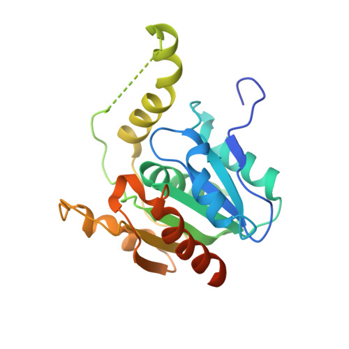
2CBG

Crystal structure of the PMSF-inhibited thioesterase domain of the fengycin biosynthesis cluster


Experimental Data Snapshot

  • Method: X-RAY DIFFRACTION
  • Resolution: 2.50 Å
  • R-Value Free: 0.237 
  • R-Value Work: 0.162 
  • R-Value Observed: 0.166 

Starting Model: experimental
View more details

wwPDB Validation   3D Report Full Report


Ligand Structure Quality Assessment 


This is version 1.5 of the entry. See complete history


Literature

The Thioesterase Domain of the Fengycin Biosynthesis Cluster: A Structural Base for the Macrocyclization of a Non-Ribosomal Lipopeptide

Samel, S.Wagner, B.Marahiel, M.A.Essen, L.-O.

(2006) J Mol Biol 359: 876

  • DOI: https://doi.org/10.1016/j.jmb.2006.03.062
  • Primary Citation of Related Structures:  
    2CB9, 2CBG

  • PubMed Abstract: 

    Many secondary metabolic peptides from bacteria and fungi are produced by non-ribosomal peptide synthetases (NRPS) where the final step of biosynthesis is often catalysed by designated thioesterase domains. Here, we report the 1.8A crystal structure of the fengycin thioesterase (FenTE) from Bacillus subtilis F29-3, which catalyses the regio- and stereoselective release and macrocyclization of the antibiotic fengycin from the NRPS template. A structure of the PMSF-inactivated FenTE domain suggests the location of the oxyanion hole and the binding site of the C-terminal residue l-Ile11 of the lipopeptide. Using a combination of docking, molecular dynamics simulations and in vitro activity assays, a model of the FenTE-fengycin complex was derived in which peptide cyclization requires strategic interactions with residues lining the active site canyon.


  • Organizational Affiliation

    Department of Chemistry, Philipps-Universität, Hans-Meerwein-Strasse, D-35032 Marburg, Germany.


Macromolecules
Find similar proteins by:  (by identity cutoff)  |  3D Structure
Entity ID: 1
MoleculeChains Sequence LengthOrganismDetailsImage
FENGYCIN SYNTHETASE244Bacillus subtilisMutation(s): 0 
UniProt
Find proteins for Q45563 (Bacillus subtilis)
Explore Q45563 
Go to UniProtKB:  Q45563
Entity Groups  
Sequence Clusters30% Identity50% Identity70% Identity90% Identity95% Identity100% Identity
UniProt GroupQ45563
Sequence Annotations
Expand
  • Reference Sequence
Small Molecules
Ligands 1 Unique
IDChains Name / Formula / InChI Key2D Diagram3D Interactions
PMS
Query on PMS

Download Ideal Coordinates CCD File 
B [auth A]phenylmethanesulfonic acid
C7 H8 O3 S
NIXKBAZVOQAHGC-UHFFFAOYSA-N
Experimental Data & Validation

Experimental Data

  • Method: X-RAY DIFFRACTION
  • Resolution: 2.50 Å
  • R-Value Free: 0.237 
  • R-Value Work: 0.162 
  • R-Value Observed: 0.166 
  • Space Group: P 61
Unit Cell:
Length ( Å )Angle ( ˚ )
a = 72.354α = 90
b = 72.354β = 90
c = 96.333γ = 120
Software Package:
Software NamePurpose
REFMACrefinement
DENZOdata reduction
SCALEPACKdata scaling
MOLREPphasing

Structure Validation

View Full Validation Report


Ligand Structure Quality Assessment 


Entry History 

Deposition Data

Revision History  (Full details and data files)

  • Version 1.0: 2007-03-06
    Type: Initial release
  • Version 1.1: 2011-05-08
    Changes: Version format compliance
  • Version 1.2: 2011-07-13
    Changes: Version format compliance
  • Version 1.3: 2017-07-05
    Changes: Data collection
  • Version 1.4: 2023-12-13
    Changes: Data collection, Database references, Derived calculations, Other, Refinement description
  • Version 1.5: 2024-11-06
    Changes: Structure summary