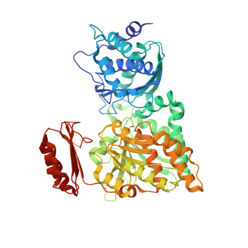
Crystal Structures of N-Acetylglucosamine-phosphate Mutase, a Member of the {alpha}-D-Phosphohexomutase Superfamily, and Its Substrate and Product Complexes.
Nishitani, Y., Maruyama, D., Nonaka, T., Kita, A., Fukami, T.A., Mio, T., Yamada-Okabe, H., Yamada-Okabe, T., Miki, K.(2006) J Biol Chem 281: 19740-19747
- PubMed: 16651269
- DOI: https://doi.org/10.1074/jbc.M600801200
- Primary Citation of Related Structures:
2DKA, 2DKC, 2DKD - PubMed Abstract:
N-acetylglucosamine-phosphate mutase (AGM1) is an essential enzyme in the synthetic process of UDP-N-acetylglucosamine (UDP-GlcNAc). UDP-GlcNAc is a UDP sugar that serves as a biosynthetic precursor of glycoproteins, mucopolysaccharides, and the cell wall of bacteria. Thus, a specific inhibitor of AGM1 from pathogenetic fungi could be a new candidate for an antifungal reagent that inhibits cell wall synthesis. AGM1 catalyzes the conversion of N-acetylglucosamine 6-phosphate (GlcNAc-6-P) into N-acetylglucosamine 1-phosphate (GlcNAc-1-P). This enzyme is a member of the alpha-D-phosphohexomutase superfamily, which catalyzes the intramolecular phosphoryl transfer of sugar substrates. Here we report the crystal structures of AGM1 from Candida albicans for the first time, both in the apoform and in the complex forms with the substrate and the product, and discuss its catalytic mechanism. The structure of AGM1 consists of four domains, of which three domains have essentially the same fold. The overall structure is similar to those of phosphohexomutases; however, there are two additional beta-strands in domain 4, and a circular permutation occurs in domain 1. The catalytic cleft is formed by four loops from each domain. The N-acetyl group of the substrate is recognized by Val-370 and Asn-389 in domain 3, from which the substrate specificity arises. By comparing the substrate and product complexes, it is suggested that the substrate rotates about 180 degrees on the axis linking C-4 and the midpoint of the C-5-O-5 bond in the reaction.
Organizational Affiliation:
Department of Chemistry, Graduate School of Science, Kyoto University, Sakyo-ku, Kyoto 606-8502, Japan.


