2DPT

Leucyl/phenylalanyl-tRNA-protein transferase complexed with puromycin


Experimental Data Snapshot

  • Method: X-RAY DIFFRACTION
  • Resolution: 2.75 Å
  • R-Value Free: 
    0.263 (Depositor), 0.250 (DCC) 
  • R-Value Work: 
    0.212 (Depositor), 0.200 (DCC) 
  • R-Value Observed: 
    0.212 (Depositor) 

Starting Model: experimental
View more details

wwPDB Validation   3D Report Full Report


Ligand Structure Quality Assessment 

Created with Raphaël 2.3.0Worse 01 BetterLigand structure goodness of fit to experimental dataBest fitted PUYClick on this verticalbar to view details

This is version 1.3 of the entry. See complete history


Literature

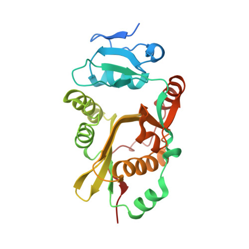
Crystal structures of leucyl/phenylalanyl-tRNA-protein transferase and its complex with an aminoacyl-tRNA analog

Suto, K.Shimizu, Y.Watanabe, K.Ueda, T.Fukai, S.Nureki, O.Tomita, K.

(2006) EMBO J 25: 5942-5950

  • DOI: https://doi.org/10.1038/sj.emboj.7601433
  • Primary Citation of Related Structures:  
    2DPS, 2DPT

  • PubMed Abstract: 

    Eubacterial leucyl/phenylalanyl-tRNA protein transferase (L/F-transferase), encoded by the aat gene, conjugates leucine or phenylalanine to the N-terminal Arg or Lys residue of proteins, using Leu-tRNA(Leu) or Phe-tRNA(Phe) as a substrate. The resulting N-terminal Leu or Phe acts as a degradation signal for the ClpS-ClpAP-mediated N-end rule protein degradation pathway. Here, we present the crystal structures of Escherichia coli L/F-transferase and its complex with an aminoacyl-tRNA analog, puromycin. The C-terminal domain of L/F-transferase consists of the GCN5-related N-acetyltransferase fold, commonly observed in the acetyltransferase superfamily. The p-methoxybenzyl group of puromycin, corresponding to the side chain of Leu or Phe of Leu-tRNA(Leu) or Phe-tRNA(Phe), is accommodated in a highly hydrophobic pocket, with a shape and size suitable for hydrophobic amino-acid residues lacking a branched beta-carbon, such as leucine and phenylalanine. Structure-based mutagenesis of L/F-transferase revealed its substrate specificity. Furthermore, we present a model of the L/F-transferase complex with tRNA and substrate proteins bearing an N-terminal Arg or Lys.


  • Organizational Affiliation

    Institute for Biological Resources and Functions, National Institute of Advanced Industrial Science and Technology, 1-1-1 Higashi, Tsukuba, Ibaraki 305-8566, Japan.


Macromolecules
Find similar proteins by:  (by identity cutoff)  |  3D Structure
Entity ID: 1
MoleculeChains Sequence LengthOrganismDetailsImage
Leucyl/phenylalanyl-tRNA--protein transferase
A, B
254Escherichia coliMutation(s): 0 
EC: 2.3.2.6
UniProt
Find proteins for P0A8P1 (Escherichia coli (strain K12))
Explore P0A8P1 
Go to UniProtKB:  P0A8P1
Entity Groups  
Sequence Clusters30% Identity50% Identity70% Identity90% Identity95% Identity100% Identity
UniProt GroupP0A8P1
Sequence Annotations
Expand
  • Reference Sequence
Experimental Data & Validation

Experimental Data

  • Method: X-RAY DIFFRACTION
  • Resolution: 2.75 Å
  • R-Value Free:  0.263 (Depositor), 0.250 (DCC) 
  • R-Value Work:  0.212 (Depositor), 0.200 (DCC) 
  • R-Value Observed: 0.212 (Depositor) 
Space Group: P 21 21 2
Unit Cell:
Length ( Å )Angle ( ˚ )
a = 115.662α = 90
b = 129.139β = 90
c = 38.571γ = 90
Software Package:
Software NamePurpose
CNSrefinement
HKL-2000data reduction
HKL-2000data scaling
CNSphasing

Structure Validation

View Full Validation Report


Ligand Structure Quality Assessment 

Created with Raphaël 2.3.0Worse 01 BetterLigand structure goodness of fit to experimental dataBest fitted PUYClick on this verticalbar to view details

Entry History 

Deposition Data

Revision History  (Full details and data files)

  • Version 1.0: 2007-01-02
    Type: Initial release
  • Version 1.1: 2008-04-30
    Changes: Version format compliance
  • Version 1.2: 2011-07-13
    Changes: Version format compliance
  • Version 1.3: 2023-10-25
    Changes: Data collection, Database references, Derived calculations, Refinement description