2ERB | pdb_00002erb

AgamOBP1, and odorant binding protein from Anopheles gambiae complexed with PEG


Experimental Data Snapshot

  • Method: X-RAY DIFFRACTION
  • Resolution: 1.50 Å
  • R-Value Free: 
    0.204 (Depositor), 0.202 (DCC) 
  • R-Value Work: 
    0.172 (Depositor), 0.170 (DCC) 
  • R-Value Observed: 
    0.172 (Depositor) 

Starting Model: experimental
View more details

wwPDB Validation 3D Report Full Report

Validation slider image for 2ERB

This is version 1.4 of the entry. See complete history

Literature

The crystal structure of an odorant binding protein from Anopheles gambiae: Evidence for a common ligand release mechanism.

Wogulis, M.Morgan, T.Ishida, Y.Leal, W.S.Wilson, D.K.

(2006) Biochem Biophys Res Commun 339: 157-164

  • DOI: https://doi.org/10.1016/j.bbrc.2005.10.191
  • Primary Citation Related Structures: 
    2ERB

  • PubMed Abstract: 

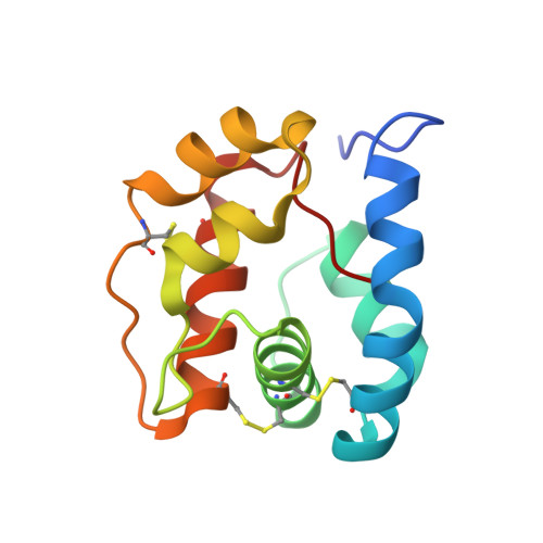
    The Anopheles gambiae mosquito is the main vector of malaria transmission in sub-Saharan Africa. We present here a 1.5A crystal structure of AgamOBP1, an odorant binding protein (OBP) from the A. gambiae mosquito. The protein crystallized as a dimer with a unique binding pocket consisting of a continuous tunnel running through both subunits of the dimer and occupied by a PEG molecule. We demonstrate that AgamOBP1 undergoes a pH dependent conformational change that is associated with reduced ligand binding. A predominance of acid-labile hydrogen bonds involving the C-terminal loop suggests a mechanism in which a drop in pH causes C-terminal loop to open, leaving the binding tunnel solvent exposed, thereby lowering binding affinity for ligand. Because proteins from two distantly related insects also undergo a pH dependent conformational change involving the C-terminus that is associated with reduced ligand affinity, our results suggest a common mechanism for OBP activity.


  • Organizational Affiliation
    • Section of Molecular and Cellular Biology, University of California, Davis, CA 95616, USA.

Macromolecule Content 

  • Total Structure Weight: 30.34 kDa 
  • Atom Count: 2,530 
  • Modeled Residue Count: 246 
  • Deposited Residue Count: 250 
  • Unique protein chains: 1

Macromolecules

Find similar proteins by:|  3D Structure
Entity ID: 1
MoleculeChains  Sequence LengthOrganismDetailsImage
odorant binding protein
A, B
125Anopheles gambiaeMutation(s): 0 

Small Molecules

Ligands 2 Unique
IDChains Name / Formula / InChI Key2D Diagram3D Interactions
PEU

Query on PEU


Download:Ideal Coordinates CCD File
D [auth B]2,5,8,11,14,17,20,23,26,29,32,35,38,41,44,47,50,53,56,59,62,65,68,71,74,77,80-HEPTACOSAOXADOOCTACONTAN-82-OL
C55 H112 O28
ISGUIIHZEJGUGQ-UHFFFAOYSA-N
MG

Query on MG


Download:Ideal Coordinates CCD File
C [auth A]MAGNESIUM ION
Mg
JLVVSXFLKOJNIY-UHFFFAOYSA-N

Experimental Data & Validation

Experimental Data

  • Method: X-RAY DIFFRACTION
  • Resolution: 1.50 Å
  • R-Value Free:  0.204 (Depositor), 0.202 (DCC) 
  • R-Value Work:  0.172 (Depositor), 0.170 (DCC) 
  • R-Value Observed: 0.172 (Depositor) 
Space Group: P 1 21 1
Unit Cell:
Length ( Å )Angle ( ˚ )
a = 30.1α = 90
b = 68.21β = 99.77
c = 61.48γ = 90
Software Package:
Software NamePurpose
CNSrefinement
MOSFLMdata reduction
CCP4data scaling
CNSphasing

Structure Validation

View Full Validation Report


Entry History 

Deposition Data

Revision History  (Full details and data files)

  • Version 1.0: 2005-12-13
    Type: Initial release
  • Version 1.1: 2008-05-01
    Changes: Version format compliance
  • Version 1.2: 2011-07-13
    Changes: Version format compliance
  • Version 1.3: 2024-04-03
    Changes: Data collection, Database references, Derived calculations, Refinement description
  • Version 1.4: 2024-11-20
    Changes: Structure summary