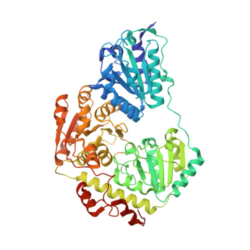
2EZT | pdb_00002ezt

Pyruvate oxidase variant F479W in complex with reaction intermediate 2-hydroxyethyl-thiamin diphosphate


Experimental Data Snapshot

  • Method: X-RAY DIFFRACTION
  • Resolution: 2.29 Å
  • R-Value Free: 
    0.187 (Depositor), 0.190 (DCC) 
  • R-Value Work: 
    0.150 (Depositor), 0.150 (DCC) 
  • R-Value Observed: 
    0.151 (Depositor) 

Starting Model: experimental
View more details

wwPDB Validation   3D Report Full Report


Ligand Structure Quality Assessment 

Created with Raphaël 2.3.0Worse 01 BetterLigand structure goodness of fit to experimental dataBest fitted FADClick on this verticalbar to view detailsBest fitted TDMClick on this verticalbar to view details

This is version 2.0 of the entry. See complete history


Literature

The catalytic cycle of a thiamin diphosphate enzyme examined by cryocrystallography.

Wille, G.Meyer, D.Steinmetz, A.Hinze, E.Golbik, R.Tittmann, K.

(2006) Nat Chem Biol 2: 324-328

  • DOI: https://doi.org/10.1038/nchembio788
  • Primary Citation of Related Structures:  
    2EZ4, 2EZ8, 2EZ9, 2EZT, 2EZU

  • PubMed Abstract: 

    Enzymes that use the cofactor thiamin diphosphate (ThDP, 1), the biologically active form of vitamin B(1), are involved in numerous metabolic pathways in all organisms. Although a theory of the cofactor's underlying reaction mechanism has been established over the last five decades, the three-dimensional structures of most major reaction intermediates of ThDP enzymes have remained elusive. Here, we report the X-ray structures of key intermediates in the oxidative decarboxylation of pyruvate, a central reaction in carbon metabolism catalyzed by the ThDP- and flavin-dependent enzyme pyruvate oxidase (POX)3 from Lactobacillus plantarum. The structures of 2-lactyl-ThDP (LThDP, 2) and its stable phosphonate analog, of 2-hydroxyethyl-ThDP (HEThDP, 3) enamine and of 2-acetyl-ThDP (AcThDP, 4; all shown bound to the enzyme's active site) provide profound insights into the chemical mechanisms and the stereochemical course of thiamin catalysis. These snapshots also suggest a mechanism for a phosphate-linked acyl transfer coupled to electron transfer in a radical reaction of pyruvate oxidase.


  • Organizational Affiliation

    Institut für Biochemie, Martin-Luther-Universität Halle-Wittenberg, Kurt-Mothes-Str. 3, 06120 Halle/Saale, Germany. georg.wille@biochemtech.uni-halle.de


Macromolecules
Find similar proteins by:  (by identity cutoff)  |  3D Structure
Entity ID: 1
MoleculeChains Sequence LengthOrganismDetailsImage
Pyruvate oxidase
A, B
603Lactiplantibacillus plantarumMutation(s): 1 
Gene Names: pox5
EC: 1.2.3.3
UniProt
Find proteins for P37063 (Lactiplantibacillus plantarum (strain ATCC BAA-793 / NCIMB 8826 / WCFS1))
Explore P37063 
Go to UniProtKB:  P37063
Entity Groups  
Sequence Clusters30% Identity50% Identity70% Identity90% Identity95% Identity100% Identity
UniProt GroupP37063
Sequence Annotations
Expand
  • Reference Sequence
Small Molecules
Ligands 6 Unique
IDChains Name / Formula / InChI Key2D Diagram3D Interactions
FAD
Query on FAD

Download Ideal Coordinates CCD File 
G [auth A],
M [auth B]
FLAVIN-ADENINE DINUCLEOTIDE
C27 H33 N9 O15 P2
VWWQXMAJTJZDQX-UYBVJOGSSA-N
TDM
Query on TDM

Download Ideal Coordinates CCD File 
F [auth A],
L [auth B]
2-[(2E)-3-[(4-AMINO-2-METHYLPYRIMIDIN-5-YL)METHYL]-2-(1-HYDROXYETHYLIDENE)-4-METHYL-2,3-DIHYDRO-1,3-THIAZOL-5-YL]ETHYL TRIHYDROGEN DIPHOSPHATE
C14 H22 N4 O8 P2 S
ZZLCKFKIIVXBQU-NTEUORMPSA-N
PO4
Query on PO4

Download Ideal Coordinates CCD File 
D [auth A],
K [auth B]
PHOSPHATE ION
O4 P
NBIIXXVUZAFLBC-UHFFFAOYSA-K
PYR
Query on PYR

Download Ideal Coordinates CCD File 
H [auth A],
N [auth B]
PYRUVIC ACID
C3 H4 O3
LCTONWCANYUPML-UHFFFAOYSA-N
MG
Query on MG

Download Ideal Coordinates CCD File 
C [auth A],
J [auth B]
MAGNESIUM ION
Mg
JLVVSXFLKOJNIY-UHFFFAOYSA-N
NA
Query on NA

Download Ideal Coordinates CCD File 
E [auth A],
I [auth B]
SODIUM ION
Na
FKNQFGJONOIPTF-UHFFFAOYSA-N
Experimental Data & Validation

Experimental Data

  • Method: X-RAY DIFFRACTION
  • Resolution: 2.29 Å
  • R-Value Free:  0.187 (Depositor), 0.190 (DCC) 
  • R-Value Work:  0.150 (Depositor), 0.150 (DCC) 
  • R-Value Observed: 0.151 (Depositor) 
Space Group: C 2 2 21
Unit Cell:
Length ( Å )Angle ( ˚ )
a = 118.79α = 90
b = 154.022β = 90
c = 165.32γ = 90
Software Package:
Software NamePurpose
DENZOdata reduction
SCALEPACKdata scaling
REFMACrefinement
PDB_EXTRACTdata extraction
MOLREPphasing

Structure Validation

View Full Validation Report


Ligand Structure Quality Assessment 

Created with Raphaël 2.3.0Worse 01 BetterLigand structure goodness of fit to experimental dataBest fitted FADClick on this verticalbar to view detailsBest fitted TDMClick on this verticalbar to view details

Entry History 

Deposition Data

Revision History  (Full details and data files)

  • Version 1.0: 2006-04-25
    Type: Initial release
  • Version 1.1: 2008-05-01
    Changes: Version format compliance
  • Version 1.2: 2011-07-13
    Changes: Derived calculations, Version format compliance
  • Version 1.3: 2017-10-18
    Changes: Refinement description
  • Version 1.4: 2021-10-20
    Changes: Data collection, Database references, Derived calculations
  • Version 1.5: 2023-08-23
    Changes: Data collection, Refinement description
  • Version 2.0: 2023-11-15
    Changes: Atomic model, Data collection