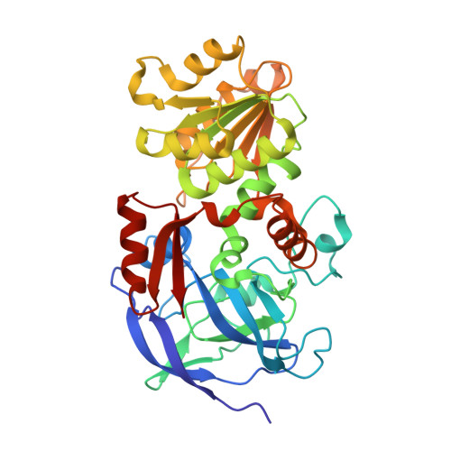
2FZW

Structure of the binary complex of the E67L mutant of human glutathione-dependent formaldehyde dehydrogenase with NAD(H)


Experimental Data Snapshot

  • Method: X-RAY DIFFRACTION
  • Resolution: 1.84 Å
  • R-Value Free: 0.204 
  • R-Value Work: 0.177 

wwPDB Validation   3D Report Full Report


Ligand Structure Quality Assessment 


This is version 1.5 of the entry. See complete history


Literature

Structure-function relationships in human glutathione-dependent formaldehyde dehydrogenase. Role of Glu-67 and Arg-368 in the catalytic mechanism.

Sanghani, P.C.Davis, W.I.Zhai, L.Robinson, H.

(2006) Biochemistry 45: 4819-4830

  • DOI: https://doi.org/10.1021/bi052554q
  • Primary Citation of Related Structures:  
    2FZE, 2FZW

  • PubMed Abstract: 

    The active-site zinc in human glutathione-dependent formaldehyde dehydrogenase (FDH) undergoes coenzyme-induced displacement and transient coordination to a highly conserved glutamate residue (Glu-67) during the catalytic cycle. The role of this transient coordination of the active-site zinc to Glu-67 in the FDH catalytic cycle and the associated coenzyme interactions were investigated by studying enzymes in which Glu-67 and Arg-368 were substituted with Leu. Structures of FDH.adenosine 5'-diphosphate ribose (ADP-ribose) and E67L.NAD(H) binary complexes were determined. Steady-state kinetics, isotope effects, and presteady-state analysis of the E67L enzyme show that Glu-67 is critical for capturing the substrates for catalysis. The catalytic efficiency (V/K(m)) of the E67L enzyme in reactions involving S-nitrosoglutathione (GSNO), S-hydroxymethylglutathione (HMGSH) and 12-hydroxydodecanoic acid (12-HDDA) were 25 000-, 3000-, and 180-fold lower, respectively, than for the wild-type enzyme. The large decrease in the efficiency of capturing GSNO and HMGSH by the E67L enzyme results mainly because of the impaired binding of these substrates to the mutant enzyme. In the case of 12-HDDA, a decrease in the rate of hydride transfer is the major factor responsible for the reduction in the efficiency of its capture for catalysis by the E67L enzyme. Binding of the coenzyme is not affected by the Glu-67 substitution. A partial displacement of the active-site zinc in the FDH.ADP-ribose binary complex indicates that the disruption of the interaction between Glu-67 and Arg-368 is involved in the displacement of active-site zinc. Kinetic studies with the R368L enzyme show that the predominant role of Arg-368 is in the binding of the coenzyme. An isomerization of the ternary complex before hydride transfer is detected in the kinetic pathway of HMGSH. Steps involved in the binding of the coenzyme to the FDH active site are also discerned from the unique conformation of the coenzyme in one of the subunits of the E67L.NAD(H) binary complex.


  • Organizational Affiliation

    Department of Biochemistry and Molecular Biology, Indiana University School of Medicine, Indianapolis, Indiana 46202-2111, USA. psanghan@iupui.edu


Macromolecules
Find similar proteins by:  (by identity cutoff)  |  3D Structure
Entity ID: 1
MoleculeChains Sequence LengthOrganismDetailsImage
Alcohol dehydrogenase class III chi chain
A, B
373Homo sapiensMutation(s): 1 
Gene Names: ADH5ADHXFDH
EC: 1.1.1.1 (PDB Primary Data), 1.1.1.284 (PDB Primary Data), 1.1.1 (UniProt)
UniProt & NIH Common Fund Data Resources
Find proteins for P11766 (Homo sapiens)
Explore P11766 
Go to UniProtKB:  P11766
PHAROS:  P11766
GTEx:  ENSG00000197894 
Entity Groups  
Sequence Clusters30% Identity50% Identity70% Identity90% Identity95% Identity100% Identity
UniProt GroupP11766
Sequence Annotations
Expand
  • Reference Sequence
Small Molecules
Ligands 4 Unique
IDChains Name / Formula / InChI Key2D Diagram3D Interactions
NAD
Query on NAD

Download Ideal Coordinates CCD File 
G [auth A],
M [auth B]
NICOTINAMIDE-ADENINE-DINUCLEOTIDE
C21 H27 N7 O14 P2
BAWFJGJZGIEFAR-NNYOXOHSSA-N
PO4
Query on PO4

Download Ideal Coordinates CCD File 
E [auth A],
J [auth B],
K [auth B]
PHOSPHATE ION
O4 P
NBIIXXVUZAFLBC-UHFFFAOYSA-K
ZN
Query on ZN

Download Ideal Coordinates CCD File 
C [auth A],
D [auth A],
H [auth B],
I [auth B]
ZINC ION
Zn
PTFCDOFLOPIGGS-UHFFFAOYSA-N
K
Query on K

Download Ideal Coordinates CCD File 
F [auth A],
L [auth B]
POTASSIUM ION
K
NPYPAHLBTDXSSS-UHFFFAOYSA-N
Experimental Data & Validation

Experimental Data

  • Method: X-RAY DIFFRACTION
  • Resolution: 1.84 Å
  • R-Value Free: 0.204 
  • R-Value Work: 0.177 
  • Space Group: P 43 21 2
Unit Cell:
Length ( Å )Angle ( ˚ )
a = 79.27α = 90
b = 79.27β = 90
c = 311.68γ = 90
Software Package:
Software NamePurpose
DENZOdata reduction
SCALEPACKdata scaling
CNSrefinement
PDB_EXTRACTdata extraction
HKL-2000data reduction
CNSphasing

Structure Validation

View Full Validation Report


Ligand Structure Quality Assessment 


Entry History 

Deposition Data

Revision History  (Full details and data files)

  • Version 1.0: 2006-06-13
    Type: Initial release
  • Version 1.1: 2008-05-01
    Changes: Version format compliance
  • Version 1.2: 2011-07-13
    Changes: Version format compliance
  • Version 1.3: 2017-10-18
    Changes: Refinement description
  • Version 1.4: 2021-10-20
    Changes: Database references, Derived calculations
  • Version 1.5: 2024-02-14
    Changes: Data collection