2G24 | pdb_00002g24

Ketopiperazine-Based Renin Inhibitors: Optimization of the "C" Ring


Experimental Data Snapshot

  • Method: X-RAY DIFFRACTION
  • Resolution: 1.90 Å
  • R-Value Free: 
    0.270 (Depositor) 
  • R-Value Work: 
    0.240 (Depositor), 0.245 (DCC) 
  • R-Value Observed: 
    0.240 (Depositor) 

wwPDB Validation 3D Report Full Report

Validation slider image for 2G24

Ligand Structure Quality Assessment 


This is version 1.3 of the entry. See complete history

Literature

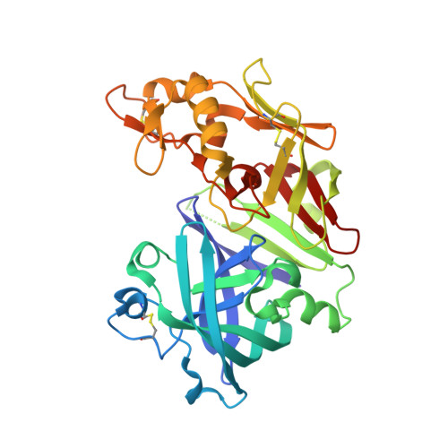
Ketopiperazine-Based Renin Inhibitors: Optimization of the C Ring

Holsworth, D.D.Cai, C.Cheng, X.M.Cody, W.L.Downing, D.M.Erasga, N.Lee, C.Powell, N.A.Ednunds, J.J.Stier, M.Jalaie, M.Zhang, E.McConnell, P.Ryan, M.J.Bryant, J.Li, T.Kasani, A.Hall, E.Subedi, R.Rahim, M.Maiti, S.

(2006) Bioorg Med Chem Lett 16: 2500-2504

  • DOI: https://doi.org/10.1016/j.bmcl.2006.01.084
  • Primary Citation Related Structures: 
    2FS4, 2G1N, 2G1O, 2G1R, 2G1S, 2G1Y, 2G20, 2G21, 2G22, 2G24, 2G26, 2G27

  • PubMed Abstract: 

    A systematic investigation of the S3 sub-pocket activity requirements was conducted. It was observed that linear and sterically small side chain substituents are preferred in the S3 sub-pocket for optimal renin inhibition. Polar groups in the S3-sub-pocket were not well tolerated and caused a reduction in renin inhibitory activity. Further, compounds with clog P's < or = 3 demonstrated a dramatic reduction in CYP3A4 inhibitory activity.


  • Organizational Affiliation
    • Pfizer Global Research and Development, Michigan Laboratories, Department of Chemistry, Ann Arbor, MI 48105, USA. Daniel.Holsworth@pfizer.com

Macromolecule Content 

  • Total Structure Weight: 73.49 kDa 
  • Atom Count: 5,214 
  • Modeled Residue Count: 658 
  • Deposited Residue Count: 666 
  • Unique protein chains: 1

Macromolecules

Find similar proteins by:|  3D Structure
Entity ID: 1
MoleculeChains  Sequence LengthOrganismDetailsImage
Renin
A, B
333Homo sapiensMutation(s): 0 
Gene Names: REN
EC: 3.4.23.15
UniProt & NIH Common Fund Data Resources
Find proteins for P00797 (Homo sapiens)
Explore P00797 
Go to UniProtKB:  P00797
PHAROS:  P00797
GTEx:  ENSG00000143839 
Entity Groups
Sequence Clusters30% Identity50% Identity70% Identity90% Identity95% Identity100% Identity
UniProt GroupP00797
Sequence Annotations
Expand
Reference Sequence

Small Molecules

Ligands 1 Unique
IDChains Name / Formula / InChI Key2D Diagram3D Interactions
7IG

Query on 7IG


Download:Ideal Coordinates CCD File
C [auth A]5-{4-[(3,5-DIFLUOROBENZYL)AMINO]PHENYL}-6-ETHYLPYRIMIDINE-2,4-DIAMINE
C19 H19 F2 N5
SRRWXMSVQYQCRX-UHFFFAOYSA-N

Experimental Data & Validation

Experimental Data

  • Method: X-RAY DIFFRACTION
  • Resolution: 1.90 Å
  • R-Value Free:  0.270 (Depositor) 
  • R-Value Work:  0.240 (Depositor), 0.245 (DCC) 
  • R-Value Observed: 0.240 (Depositor) 
Space Group: P 21 3
Unit Cell:
Length ( Å )Angle ( ˚ )
a = 140.885α = 90
b = 140.885β = 90
c = 140.885γ = 90
Software Package:
Software NamePurpose
CNSrefinement

Structure Validation

View Full Validation Report


Ligand Structure Quality Assessment 


Entry History 

Deposition Data

Revision History  (Full details and data files)

  • Version 1.0: 2006-06-13
    Type: Initial release
  • Version 1.1: 2008-05-01
    Changes: Version format compliance
  • Version 1.2: 2011-07-13
    Changes: Version format compliance
  • Version 1.3: 2024-11-20
    Changes: Data collection, Database references, Derived calculations, Structure summary