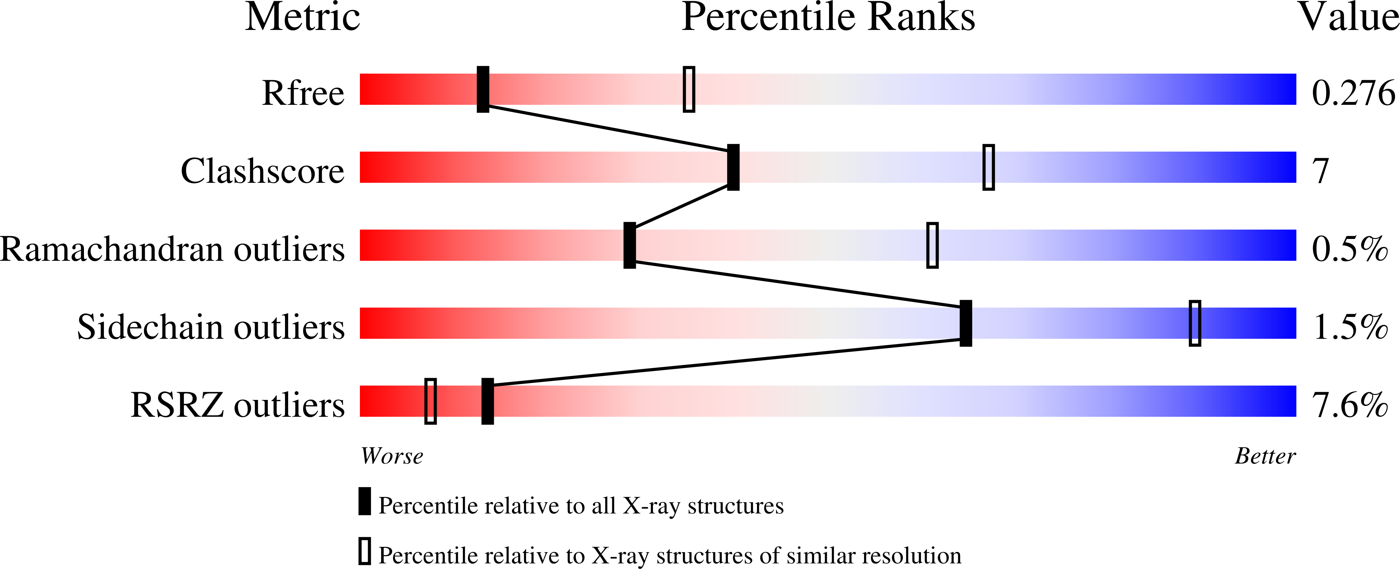
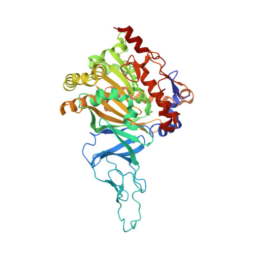
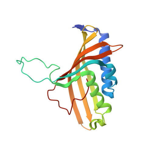
2GBX

Crystal Structure of Biphenyl 2,3-Dioxygenase from Sphingomonas yanoikuyae B1 Bound to Biphenyl


Experimental Data Snapshot

  • Method: X-RAY DIFFRACTION
  • Resolution: 2.80 Å
  • R-Value Free: 0.268 
  • R-Value Work: 0.237 
  • R-Value Observed: 0.238 

Starting Model: experimental
View more details

wwPDB Validation   3D Report Full Report


Ligand Structure Quality Assessment 


This is version 1.4 of the entry. See complete history


Literature

Structural investigations of the ferredoxin and terminal oxygenase components of the biphenyl 2,3-dioxygenase from Sphingobium yanoikuyae B1.

Ferraro, D.J.Brown, E.N.Yu, C.L.Parales, R.E.Gibson, D.T.Ramaswamy, S.

(2007) BMC Struct Biol 7: 10-10

  • DOI: https://doi.org/10.1186/1472-6807-7-10
  • Primary Citation of Related Structures:  
    2GBW, 2GBX, 2I7F

  • PubMed Abstract: 

    The initial step involved in oxidative hydroxylation of monoaromatic and polyaromatic compounds by the microorganism Sphingobium yanoikuyae strain B1 (B1), previously known as Sphingomonas yanoikuyae strain B1 and Beijerinckia sp. strain B1, is performed by a set of multiple terminal Rieske non-heme iron oxygenases. These enzymes share a single electron donor system consisting of a reductase and a ferredoxin (BPDO-FB1). One of the terminal Rieske oxygenases, biphenyl 2,3-dioxygenase (BPDO-OB1), is responsible for B1's ability to dihydroxylate large aromatic compounds, such as chrysene and benzo[a]pyrene. In this study, crystal structures of BPDO-OB1 in both native and biphenyl bound forms are described. Sequence and structural comparisons to other Rieske oxygenases show this enzyme to be most similar, with 43.5 % sequence identity, to naphthalene dioxygenase from Pseudomonas sp. strain NCIB 9816-4. While structurally similar to naphthalene 1,2-dioxygenase, the active site entrance is significantly larger than the entrance for naphthalene 1,2-dioxygenase. Differences in active site residues also allow the binding of large aromatic substrates. There are no major structural changes observed upon binding of the substrate. BPDO-FB1 has large sequence identity to other bacterial Rieske ferredoxins whose structures are known and demonstrates a high structural homology; however, differences in side chain composition and conformation around the Rieske cluster binding site are noted. This is the first structure of a Rieske oxygenase that oxidizes substrates with five aromatic rings to be reported. This ability to catalyze the oxidation of larger substrates is a result of both a larger entrance to the active site as well as the ability of the active site to accommodate larger substrates. While the biphenyl ferredoxin is structurally similar to other Rieske ferredoxins, there are distinct changes in the amino acids near the iron-sulfur cluster. Because this ferredoxin is used by multiple oxygenases present in the B1 organism, this ferredoxin-oxygenase system provides the structural platform to dissect the balance between promiscuity and selectivity in protein-protein electron transport systems.


  • Organizational Affiliation

    Department of Biochemistry, University of Iowa Roy J. and Lucille A. Carver College of Medicine, Iowa City, Iowa, 52242, USA. daniel-ferraro@uiowa.edu


Macromolecules
Find similar proteins by:  (by identity cutoff)  |  3D Structure
Entity ID: 1
MoleculeChains Sequence LengthOrganismDetailsImage
Biphenyl 2,3-Dioxygenase Alpha Subunit
A, C, E
454Sphingobium yanoikuyaeMutation(s): 0 
Gene Names: bphA1fA2f
UniProt
Find proteins for A2TC87 (Sphingobium yanoikuyae)
Explore A2TC87 
Go to UniProtKB:  A2TC87
Entity Groups  
Sequence Clusters30% Identity50% Identity70% Identity90% Identity95% Identity100% Identity
UniProt GroupA2TC87
Sequence Annotations
Expand
  • Reference Sequence
Find similar proteins by:  (by identity cutoff)  |  3D Structure
Entity ID: 2
MoleculeChains Sequence LengthOrganismDetailsImage
Biphenyl 2,3-Dioxygenase Beta Subunit
B, D, F
174Sphingobium yanoikuyaeMutation(s): 0 
UniProt
Find proteins for A2TC88 (Sphingobium yanoikuyae)
Explore A2TC88 
Go to UniProtKB:  A2TC88
Entity Groups  
Sequence Clusters30% Identity50% Identity70% Identity90% Identity95% Identity100% Identity
UniProt GroupA2TC88
Sequence Annotations
Expand
  • Reference Sequence
Small Molecules
Ligands 4 Unique
IDChains Name / Formula / InChI Key2D Diagram3D Interactions
FES
Query on FES

Download Ideal Coordinates CCD File 
H [auth A],
N [auth C],
T [auth E]
FE2/S2 (INORGANIC) CLUSTER
Fe2 S2
NIXDOXVAJZFRNF-UHFFFAOYSA-N
BNL
Query on BNL

Download Ideal Coordinates CCD File 
I [auth A],
O [auth C],
U [auth E]
BIPHENYL
C12 H10
ZUOUZKKEUPVFJK-UHFFFAOYSA-N
ZN
Query on ZN

Download Ideal Coordinates CCD File 
J [auth B]
K [auth B]
L [auth B]
P [auth D]
Q [auth D]
J [auth B],
K [auth B],
L [auth B],
P [auth D],
Q [auth D],
R [auth D],
V [auth F],
W [auth F],
X [auth F]
ZINC ION
Zn
PTFCDOFLOPIGGS-UHFFFAOYSA-N
FE
Query on FE

Download Ideal Coordinates CCD File 
G [auth A],
M [auth C],
S [auth E]
FE (III) ION
Fe
VTLYFUHAOXGGBS-UHFFFAOYSA-N
Experimental Data & Validation

Experimental Data

  • Method: X-RAY DIFFRACTION
  • Resolution: 2.80 Å
  • R-Value Free: 0.268 
  • R-Value Work: 0.237 
  • R-Value Observed: 0.238 
  • Space Group: P 31 2 1
Unit Cell:
Length ( Å )Angle ( ˚ )
a = 133.94α = 90
b = 133.94β = 90
c = 219.708γ = 120
Software Package:
Software NamePurpose
XSCALEdata scaling
AMoREphasing
REFMACrefinement
PDB_EXTRACTdata extraction
JDirectordata collection

Structure Validation

View Full Validation Report


Ligand Structure Quality Assessment 


Entry History 

Deposition Data

Revision History  (Full details and data files)

  • Version 1.0: 2007-03-20
    Type: Initial release
  • Version 1.1: 2008-05-01
    Changes: Version format compliance
  • Version 1.2: 2011-07-13
    Changes: Non-polymer description, Version format compliance
  • Version 1.3: 2017-10-18
    Changes: Refinement description
  • Version 1.4: 2023-08-30
    Changes: Data collection, Database references, Derived calculations, Refinement description