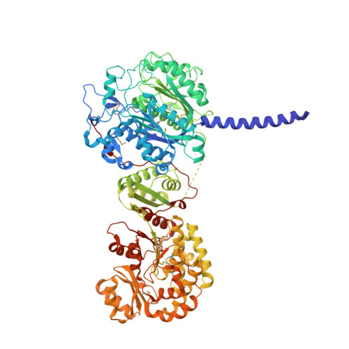
2HG4

Structure of the ketosynthase-acyltransferase didomain of module 5 from DEBS.


Experimental Data Snapshot

  • Method: X-RAY DIFFRACTION
  • Resolution: 2.73 Å
  • R-Value Free: 0.255 
  • R-Value Work: 0.216 
  • R-Value Observed: 0.218 

wwPDB Validation   3D Report Full Report


This is version 1.3 of the entry. See complete history


Literature

The 2.7-A crystal structure of a 194-kDa homodimeric fragment of the 6-deoxyerythronolide B synthase.

Tang, Y.Kim, C.Y.Mathews, I.I.Cane, D.E.Khosla, C.

(2006) Proc Natl Acad Sci U S A 103: 11124-11129

  • DOI: https://doi.org/10.1073/pnas.0601924103
  • Primary Citation of Related Structures:  
    2HG4

  • PubMed Abstract: 

    The x-ray crystal structure of a 194-kDa fragment from module 5 of the 6-deoxyerythronolide B synthase has been solved at 2.7 Angstrom resolution. Each subunit of the homodimeric protein contains a full-length ketosynthase (KS) and acyl transferase (AT) domain as well as three flanking "linkers." The linkers are structurally well defined and contribute extensively to intersubunit or interdomain interactions, frequently by means of multiple highly conserved residues. The crystal structure also reveals that the active site residue Cys-199 of the KS domain is separated from the active site residue Ser-642 of the AT domain by approximately 80 Angstrom. This distance is too large to be covered simply by alternative positioning of a statically anchored, fully extended phosphopantetheine arm of the acyl carrier protein domain from module 5. Thus, substantial domain reorganization appears necessary for the acyl carrier protein to interact successively with both the AT and the KS domains of this prototypical polyketide synthase module. The 2.7-Angstrom KS-AT structure is fully consistent with a recently reported lower resolution, 4.5-Angstrom model of fatty acid synthase structure, and emphasizes the close biochemical and structural similarity between polyketide synthase and fatty acid synthase enzymology.


  • Organizational Affiliation

    Department of Chemistry, Stanford University, Stanford, CA 94305, USA.


Macromolecules
Find similar proteins by:  (by identity cutoff)  |  3D Structure
Entity ID: 1
MoleculeChains Sequence LengthOrganismDetailsImage
6-Deoxyerythronolide B Synthase
A, B, C, D, E
A, B, C, D, E, F
917Saccharopolyspora erythraeaMutation(s): 17 
Gene Names: eryA
EC: 2.3.1.94
UniProt
Find proteins for Q03133 (Saccharopolyspora erythraea)
Explore Q03133 
Go to UniProtKB:  Q03133
Entity Groups  
Sequence Clusters30% Identity50% Identity70% Identity90% Identity95% Identity100% Identity
UniProt GroupQ03133
Sequence Annotations
Expand
  • Reference Sequence
Small Molecules
Ligands 3 Unique
IDChains Name / Formula / InChI Key2D Diagram3D Interactions
SO4
Query on SO4

Download Ideal Coordinates CCD File 
H [auth A],
J [auth B],
M [auth C],
R [auth E],
U [auth F]
SULFATE ION
O4 S
QAOWNCQODCNURD-UHFFFAOYSA-L
ACT
Query on ACT

Download Ideal Coordinates CCD File 
G [auth A]
I [auth B]
L [auth C]
O [auth D]
Q [auth E]
G [auth A],
I [auth B],
L [auth C],
O [auth D],
Q [auth E],
T [auth F]
ACETATE ION
C2 H3 O2
QTBSBXVTEAMEQO-UHFFFAOYSA-M
CL
Query on CL

Download Ideal Coordinates CCD File 
K [auth B],
N [auth C],
P [auth D],
S [auth E],
V [auth F]
CHLORIDE ION
Cl
VEXZGXHMUGYJMC-UHFFFAOYSA-M
Modified Residues  1 Unique
IDChains TypeFormula2D DiagramParent
MSE
Query on MSE
A, B, C, D, E
A, B, C, D, E, F
L-PEPTIDE LINKINGC5 H11 N O2 SeMET
Experimental Data & Validation

Experimental Data

  • Method: X-RAY DIFFRACTION
  • Resolution: 2.73 Å
  • R-Value Free: 0.255 
  • R-Value Work: 0.216 
  • R-Value Observed: 0.218 
  • Space Group: C 1 2 1
Unit Cell:
Length ( Å )Angle ( ˚ )
a = 305.258α = 90
b = 150.149β = 110.03
c = 184.378γ = 90
Software Package:
Software NamePurpose
REFMACrefinement
XDSdata reduction
XDSdata scaling
SHELXphasing
RESOLVEphasing

Structure Validation

View Full Validation Report


Entry History 

Deposition Data

Revision History  (Full details and data files)

  • Version 1.0: 2006-07-11
    Type: Initial release
  • Version 1.1: 2008-04-29
    Changes: Version format compliance
  • Version 1.2: 2011-07-13
    Changes: Advisory, Derived calculations, Version format compliance
  • Version 1.3: 2024-11-06
    Changes: Data collection, Database references, Derived calculations, Refinement description, Structure summary